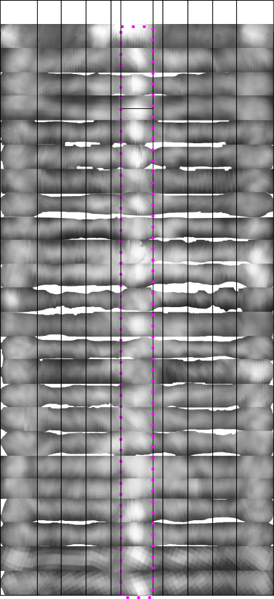
![]() Here are 24 arms from 12 SP-D dodecamers which I have cut out, straightened and placed in a vertical row, using a single width as the diminishing grayscale point on the two ends (the CRD). The vertical set on the left has width adjusted to a given size but it is clear (dotted pink rectangle) that the centers vary (Ntermini-junctins). In the 24 arms on the right column the images have been cut in the middle, and the centers “centered” which will make an overlay of the Ntermini peaks easier to measure in width. The point of this exercise (which does not use all the images I have gathered) is to test whether a composit image which has been adjusted to x nm width and central peak centered will show a definitive number of peaks between the N terminus peak and the peripheral CRD peaks.
Here are 24 arms from 12 SP-D dodecamers which I have cut out, straightened and placed in a vertical row, using a single width as the diminishing grayscale point on the two ends (the CRD). The vertical set on the left has width adjusted to a given size but it is clear (dotted pink rectangle) that the centers vary (Ntermini-junctins). In the 24 arms on the right column the images have been cut in the middle, and the centers “centered” which will make an overlay of the Ntermini peaks easier to measure in width. The point of this exercise (which does not use all the images I have gathered) is to test whether a composit image which has been adjusted to x nm width and central peak centered will show a definitive number of peaks between the N terminus peak and the peripheral CRD peaks.
I think I will try to make a case for a central peak (which is obvious) and four (which includes the CRD) peaks on each side…THe CRD peak is obviously three lumps, and the three peaks between the N and CRD are subtle, and decline in LUT values and are not all the same width.
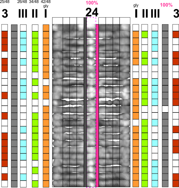 Pink=100% have N termini peaks; gray, 100% have the CRD peaks, but only half the arms have all three peaks between the former two (red). The peak considered the glycosylation site (orange), two minor peaks lateral to that (II and III) are green and blue respectively. Incidence of “eyeball” peaks declines moving outward on the arms.