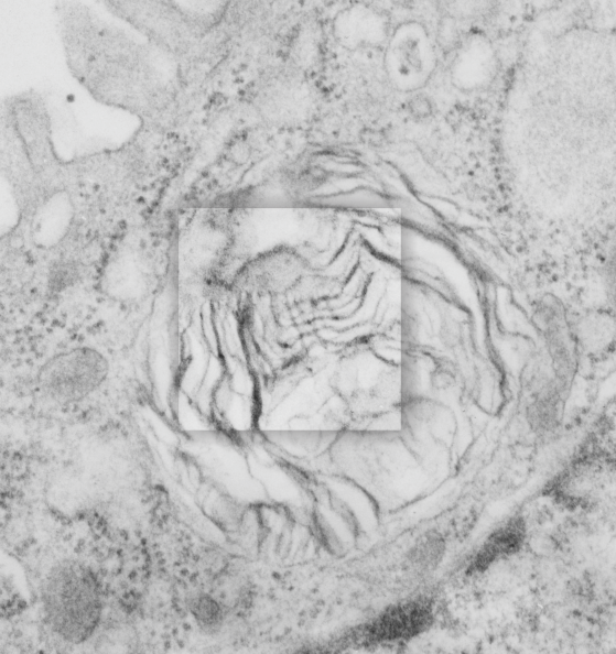
Lamellar body in an alveolar type II cell has surfactant-like-tubular-myelin grid pattern. This was found in an electron micrograph of a ferret type II cell (neg 9872, block 23462, ferret # 7, control, 6 10 1982). This particular lamellar body is unusual in that within the lamellar structure, there is a grid like pattern which is normally seen only in the alveolar space. While, according to the literature, alveolar type II cells can internalize mature surfactant from the alveolar space, this doesnt anatomically seem very likely to me as an explanation for this particular structure. I thought it was unusual enough to post. I put a bounding box and a drop shadow around the area of interest. The grid inside has at least 4 partitions horizontally as well as vertically. Surfactant protein A is likely responsible for the grid (according to consensus) though this is usually in the alveolar space. This mineature tubular myelin was described in a book, ed Jacques Bourbon I think, which was reported to be about 1/3 the size of tubular myelin in the alveolar space.
Here are two reference images for the above micrograph to give approximate sizes. Regular tubular myelin has a side to side distance of something around 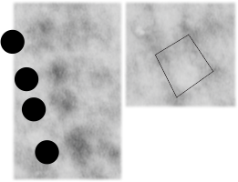 100 nm, whereas this intra-lamellar-body mineature tubular myelin sample is about 60 nm? Ribosomes are 25-30 nm (black circles)