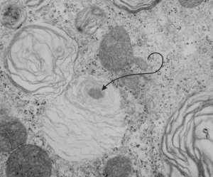
 In lung, the type II cell has lamellar bodies, the secretion granule of surfactant, but not all species show a projection core within those lamellar bodies. I don’t know if guinea pig has ever been described as having projection cores in their lamellar bodies, but this looks either like a portion of a multivesicular body-fusion with lamellar body, or maybe a projection core (which should probably have more layered substructure). It has a different electron microscopic appearance than lipid does, and lipid droplets, seen as mostly textureless round areas, are found in lamellar bodies very frequently, and in many species in my experience. Here is a crop from a type II cell from guinea pig lung and the lamellar body (end of pointer) seems to me to have a projection granule. (6481_M8005 guinea pig #51, vinyl chloride exposed plus 2mg vitamin C you can access this pdf with specifics and details of animal exposures vinyl_chloride_vit_C_guinea_pig_lung )
I also went quickly through the electron micrographs I have for dog lung (467 lamellar bodies quickly counted, with 24 lamellar bodies equivocal for a projection core, but not really a good match, looking much more like areas where there is multivesicular body material.
Also, perusing over 1000 lamellar bodies in electron micrographs from ferret my conclusion is that there are many little lipid droplets within lamellar bodies, but only a rare protein-like body that might under ideal fixation circumstances be seen as a projection body but still without substructure that is layered (which Ochs 2009 mentions is the hallmark for projection cores. He also states that they do not appear in rodents). There are many many examples of multivesicular type densities within lamellar bodies though, clearly defined and usually around the periphery of the lamellar bodies. That said, I understand that “fixation” is everything.
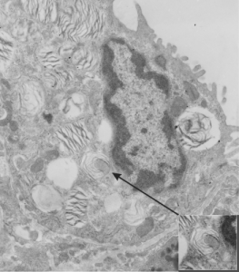
Here is an electron micrograph of ferret alveolar type II cell which has possibly as good an indication from archived photos as any that maybe in some ideal conditions, ferret might actually have some lamellar bodies with some type of density, maybe part of a multivesicular body-core together. I am not banking on it, but offer this, as one in a thousand example of a lamellar body that might have a projection core. As with guinea pig and dog, most of the roundish things at one or more poles or periphery of a lamellar body look more like lipid than layered protein. Lipid is common. This could just as easily be a tangential section of the bottom side of a multivesicular body fusing with a lamellar body. And only specific studies will determine which is which, and most likely will turn out to be something of both together.
