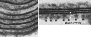
So do you think that SP-A is the main protein component of the alveolar type II cell granule? SP-B has been shown to work with SP-A in several circumstances and particularly in forming tubular myelin lattice structures. SP-B is a pretty small and flat molecule (but not as flat as DP-C, and it diagrams seem to place it wort of “surface-wise) to the plasmalemma bilayer. and if i remember right, maybe the bilayer elements of the alveolar space. Here is a cut and paste from a paper from the “before times” by Williams et al, 1991, where they show artifically created showing some lamellar ultrastructural similarities with the two dense lines and central punctate line seen in RER granules of alveolar type II cells. (but I am not sure about size) thought the mention of 20 nm is there, because i don’t know if that is a measurement from one dense lipid layer to the center punctate line, or all the way across the lamellae. Her reference is HERE. and a cropped figure from her publication is below. The match is certainly not perfect, but suggestive of possible structural organization of SP-A and lipid.
