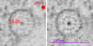
Electron micrograph of an alveolar type Ii cell which is overrun with surfactant protein granules and an isolated part of a larger granule shows this profile which is so symmetrical as to look like the spokes of a wheel. Micrograph on left is unretouched, micrograph on the right is burned in the areas of periodicity of the molecules of surfactant protein, and dodged in the areas between, just slightly to highlight what I saw.
The purpose of the study is still to determine which of the surfactant proteins is responsible for forming the granules. So here is such a single period of a granule which has a ribosome marked for approximate size (red dot=27 nm) red bar=100 nm superimposed on an electron micrograph (unretouched). Right hand image has emphasized image, and a circumference of the round less-dense inner band of the period (central dense dot represents the “outer dense band” seen in linear profiles. The inner dots have been counted in linear granules at closer to 5-6 per 100 nm but here there are 11 or 12 dots of the less dense central band under the circumference, which is about 330 nm, or a little less, and in this photo the dots measure about 15-17 nm (consistent with previous measurements, then are spaced at about 25 nm apart. Not everything lines up exactly with previous posts on this subject in the linear profiles, but clearly they are analogous and match pretty well. The spoke appearance likely corresponds to the vertical lineup of molecules seen in granules that have a linear arrangement. (just as an aside, there is something spooky about the fact that I have found several times now that the space between these molecules in the central less dense band are spaced apart the same distance as a ribosome…. that is just uncanny, and I bet it will end up meaning something about protein synthesis in general–just a heads up). see this post and this post
