Methods of fixation and staining allow ultrastructural components of each cell type to be preserved and resolved differently. Glutaraldehyde based, non-coagulant-fixatives encourage direct cross-links with amines and other nucleophiles, most notably lysine and arginine, and cause deformation of the alpha-helix structures in proteins. Paraformaldehyde cross-links basic amino acid lysine, as well. Osmium tetroxide, in these studies was always used as a secondary fixative, except for beagle dog, where it was used as a primary fixative, helps to preserve lipids, and cross-links at unsaturated sites in membrane lipids and nucleophiles like amino and sulfhydryl groups. Osmium tetroxide enhances overall electron density of the specimen, adding contrast. Contrast is further enhanced by positive staining with heavy metal salts (uranyl acetate, enhancing lipids, nucleoproteins and proteins) and lead citrate (enhancing the contrast of proteins, membranes and glycogen) (Frankl, 2015; wikipedia). These all fit well with the type of layering (electron dense banding) found in alveolar type II cell granules.
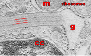
So what electron density in the structure of the alveolar type II cell reveal about the molecular structure of the layered protein. Well, surfactant protein A (and D also) has helical regions, probably hopefully the less dense central layers have a lot of lysine and sulfhydryl groups. This might fit well with surfactant protein A patterning. Three red lines = 2 outer dense layers and less dense central layer; m=mitochondrion; ribosomes are the black dots (nucleoprotein staining with uranyl acetate), especially situated on the electron lucent racket end of the granule; cc= condensed nuclear chromatin (nucleoprotein staining with uranyl acetate), in the rounded portion of nucleus in the lower left corner of the micrograph. This image was shadowed with an exact replica, 68% transparency, and a few pixels to right and lower than bottom image. one of my favorite electron micrographs of these alveolar type II cell granules .