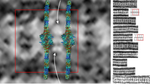
Lots of images have been published for Burbeck granules. I am interested in them only because they seem to be the only other C-type lectin which has a substructure that is organized and obvious when viewed with electron microscopy. Well not the only, because I think that SP-D has fuzzy ball structure that is seen with TEM. It is puzzling why MBP and some other lectins don’t assemble into orderly granules like langerin, and SP-A (at least I think it is SP-A).
So here is a composite that I have made from TEMs posted everywhere in the literature, lined up with the 50 nm distance between the ER membranes, and such a very predictable substructure of proteins organized within. One publication in Plos ONE (Glycosaminoglycans Are Interactants of Langerin: Comparison with gp120 Highlights an Unexpected Calcium-Independent Binding Mode. Eric Chabrol, Alessandra Nurisso, Antoine Daina, Emilie Vassal-Stermann, Michel Thepaut, Eric Girard, Romain R. Vivès, Franck Fieschi) gives a molecular structure and a hypothetical overlay onto TEM images. I think they missed it by a little…. for starters their intramembrane part of the molecule is NOT shown in their diagrams, which makes the molecular fit difficult since the CRD of the molecule are a relatively larger portion of the actual TEMs than is seen in their molecular diagram (line diagrams on the far right of the image). Additionally, there is a cytoplasmic portion of langerin, at opposite ends of the mirrored CRD portions of langerin which their diagram does not show (don’t know why they chose not to show it). I vectorized their diagram and have placed it back in the 50 nm membrane boundaries of the ER with the transmembrane portions actually existing and that makes the size ration of the model molecule fit the actual TEM much better.
Again, my only interest in this is that I am trying to ascertain whether my thought that SP-A is responsible for the granule in guinea pig and ferret type II alveolar cells true.
Right side of the diagram is a collection of Birbeck granule TEMs sized to 50 nm, the image on the right shows a red 50 nm bounding box, and four langerin molecules oriented CRD inward, and transmembrane areas through the ER membrane. Just my opinion here but there is another molecule or some portion of langerin which is “between” the four diagrams superimposed. White arrows point there. Their diagrams (two alternatives) are shown in red and grey to the right of the actual TEMs of Birbeck granules.