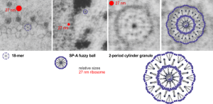
Still working on this granule in alveolar type II cells. I think there are three levels of organization to the cylindrical and circumferential manifestations of whatever surfactant protein comprises these granules. The original 18-mer, well confirmed by numerous other scientists, on the left, relative to the size of a ribosome, and also with the hexagonal order seen in tangential sections of the outer dense layer of the granule. Middle image is the proposed SP-A fuzzy ball which seems to be a spherical organization of the same 18-mer, just with something different happening in the center region of the sphere. I can’t even guess what that organization (missing perhaps some of the CRD regions of SP-A?) to make things fit. This image also relative to the size of a single ribosome (red dot). Then on the right hand side is a double-layered cylindrical granule (this one likely from guinea pig) which has a diameter of about 200 nm, which means is likely has a column of four molecules for each radius. Size of the actual superimposed cylindrical period, and then a relatively sized – diagram… sized relative to the other spherical or concentric configurations of SP-A. The micrographs do pretty much dictate the arrangements, or at least that was what dictated the arrangements of the SP-A stick figure I used (which was vectorized from models of SP-A in publications.
