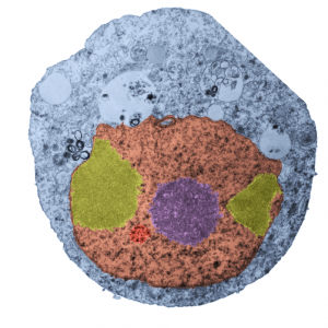
Nothing is black and white, except electron micrographs, and even then they are easy game for dodging, blending, blurring, mending, and pseudocoloring. Cytoplasm here is blue. In this case I have a cell from a culture of A549 cells in which C9orf82 was knocked down using si67. This is pellet 4 from one of those studies. It produced this cell in which the granular component of the nucleolus was separated into at least two bodies (almost bilaterally symmetrically arranged) which rest at the inner nuclear membrane. Fibrillar centers are small, dense fibrillar component is still visible (albeit not distinctly) in the nucleolus (pseudocolored purple in the center of the cell). Off to the side is what I could guess would be either degraded DNA (not my first choice as an answer (it is pseudocolored bile-yellow) or perhaps a fine granular component of the nucleolus in an apoptotic cell. Part of an interchromatin granule (red, lower left) cluster (IGC) (sometimes called speckles, a name which is not really to my liking since the IGC has a boundary, a background texture that is also part of the structure). So I would propose keeping two names, the “IGC” which is the greater boundary of that area and “nuclear speckles” as well, since the densities (as seen in this micrograph) can apparently be large.
If i am not off base here, the background of the whole IGC is red, but there are very clearly larger than typical speckles within the IGC, and the latter itself is quite small. With immunohistochemistry the diffuse staining of IGC background proteins fluoresces with some of the antibodies to proteins like SC-37 in an area which can be 2 microns across easily. I am looking for alternative proteins stained in very punctate regions of the IGC and hopefully they will be different and good markers for a separate ultrastructural components of the IGC. The point would be to localize to the densities with the IGC, some proteins for splicing, and accept that the diffuse staining could apply to SC-37, but not other proteins, mainly those in the variable size granules within the IGC.
We will see if there are data to back up the keeping of both names but assigning them to their obvious separate entities. BTW, there is pretty obvious bilateral symmetry to this apoptotic cell nucleus. and a radial symmetry to the dense bodies within the red colored IGC. And while i have no clue what the curley-Qs are… i bet they have something to do with the transfection. Just looking at this cell shows one tiny surviving mitochondrion, in this apoptotic cell, which is very close to the cytoplasmic filaments of a nuclear pore…also something which needs to be monitored during apoptosis.
