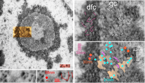
Epithelial cell in the lung of a baboon (probably a type II alveolar cell) showed a ring nucleolus with a single fibrillar center and concentric granular and dense fibrillar components. I took the time to measure (relative to a series of ribosomes measured in the adjacent cytoplasm – estimated at 27nm in diameter) the size of the granules in the dense fibrillar portion and in the granular portion of this nucleolus. The image on the left shows the whole nucleolus, the three divisions (there are probably more) of the nucleolus are prominent. From that image of the nucleolus, the orange area is enlarged twice on the right to highlight distance between granules of the dense fibrillar component and the granular component. The small redish box is what was used to estimate the size of those granules by measuring ribosomes therin (red dot) obtained from a portion of adjacent cytoplasm from the same image.
The diagonal (and rearranged and measured as horizontal) lines are those between granules in the granular portion (white) and also the dense fibrillar portion (purple). The spaces between the granules in the granular portion are marked out with a peach transparency and the blue circles represent the size of granules in that area.
Key: orange circles = size of the granules in the dense fibrillar portion at 30nm, with about 38nm distance between them; white lines and blue circles 21nm) represent the diameter of the granules in the granular portion and the distance between them (42nm). For comparison, the ribosome (red dot, lower center-left is 27 nm, and the distance between ribosomes is about 58nm. The apparently-coiled strings (pink) are something that occurred regularly, kind of an arcing gently oval beaded appearance. 13279_94-32_baboon_lung_PFP (perfluorophenanthrene).
