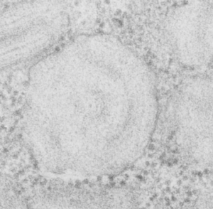
Guinea pig, aged, routine electron microscopy, type II alveolar cell, in particular the RER and a highly organized protein contained: possibly surfactant protein A.
This micrograph is one of the few that is concentric and shows banding at the same time, at least that I have encountered. The center period looks like the others which are only a single set of bands, i.e. a single central dark area, a lighter band, and then the outer band (which would include the CDR part of the protein octadecamers and the RER membrane.
It has ribosomes for size comparison, at 20-30 nm in diameter, and as can be seen from the longitudinal profile of RER in the upper left corner where the banding is more obvious, even in the concentric arrangement, the spacing of a single complete period is about 100 nm. I evened out the exposure of this micrograph, nothing else.
