Category Archives: Ultimate order, the cell

The beauty and order of life is astounding.

Anamnestic – does this describe some lichen planus variants? Random thoughts


The following paragraphs are just random thoughts and articles that might shed some light on the disease that is lichen planus (thinking too some variants of the disease like hypertrophic lichen planus and lichenoid drug-induced eruptions (which i would rename lichenoid like dermatoses) with various environmental triggers. I found a relatively old publication in the 1990s that clearly felt environmental factors were important in LP, but with passing years and changes in research-topic “runway-fashions” the environmental influence gets less attention.

Reading an article on histologic distinctions between lichen planus and lichenoid drub-induced dermatitis (link reference given previously) I think it is a perfect description of what I have experienced and a perfect expression of the gene environment interactions that go along with other cases where the immune system decides to make massive amounts of antibodies to a “wrong thing” — as the online definition of anamnestic states so beautifully “an enhanced reaction of the body’s immune system to an antigen that is related to an antigen previously encountered”

I cant wait (but am apparently going to wait a long time) for my condition to clear so i can go back and test if my assumption about mortar, grout, cement, concrete and additives are the “guilty-by-association antigens” that cause this disease.

AND here is a new tack. duh, i should have thought this up a long time ago. I have been slightly aware that potatoes (not sweet potatoes or yams) might cause an increase in itchiness of my lichen planus spots. I did cut out potatoes and peppers for a while, but then on June 6 2018 (that is about 9 months into the flare up of this disease in my) I went back to eating peppers tomatoes and potatoes (all nightshade family members). Yesterday i cut out potatoes again…. so Then i googled, nightshade plants and immune response and find again, what i knew once, that those have “lectins”. Make the connection here. Lung cells (in particular, alveolar type II cells) secrete surfactant protein A… the latter is named a surfactant protein, and it does in fact participate in the formation of tubular myelin in the alveolar spaces but one can live just fine and had sufficient lung function WITHOUT surfactant A. That said, there are immune consequences because surfactant A is a C-type lectin (lung reaction allergens) and

“Antigen-presenting cells can capture antigens that are deposited in the skin, including vaccines given subcutaneously” (Flatcher et al, J. Invest. Derm. 2010 Mar;130(3):755-62. doi: 10.1038/jid.2009.343).
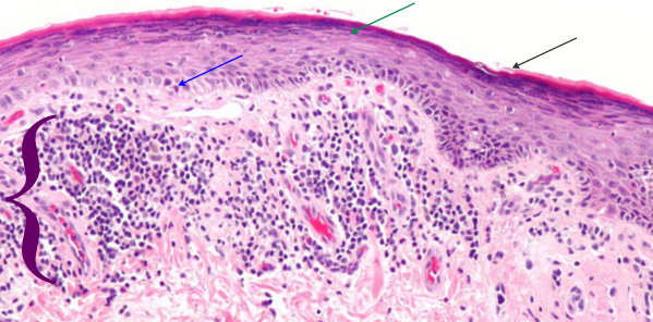
Aside: shame on whomever called lichen planus a benign situation… ha ha.. clearly never had it. Bless the person who describes the causes of lichen planus as “undefined” because clearly there is a cause, and likely a multitude of causes, and I add, many are going to be environmental… mistaken identity immunity, or inappropriate immune responses. Bless the guy who mentions adhesion molecules in this disease…. i have no clue how the extracellular adhesion molecules in epidermis can tell the difference between “sheer” (rubbing) and patting (e.g. patting dry an area of lichen planus eruptions) but the cells know… a tiny bit of sheer tension and it triggers highly intense itching. It only took one google search to find this article which actually mentions extracellular adhesion molecules in Pemphigus, so this is not far from responses in lichen planus.

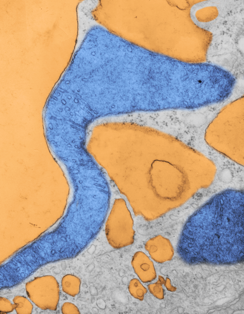
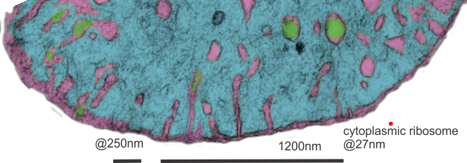
And another article here which describes roles for langerhans cells… goodness, maybe direct access of environmental materials (as in my case, cement, mortar, thinset mortar grout, and whatever was present on acorn debris thrown up into my shins from my lawnmower) was opsonized by the langerin (C-type lectin) surface protein in my epidermal langerhans cells… ha ha… so much fun to learn this stuff… and to suppose what processes might have occurred.

One observation is that LCs induce some form of contact hypersensitivity though the immunostimulatory capacity of langerhans cells depends on the quality and quantity of the environmental stimuli. “Immune responses can be markedly enhanced when antigens are ‘‘targeted’’ to uptake receptors on dendritic cells and administered together with  inflammatory stimuli.” (quoted from an article  “Changing Views of the Role of Langerhans Cells NikolausRomani, Patrick M.Brunner, GeorgStingl”
I also saw where antigens (example was arsenic) could be captured by langerhans cells directly is damaged skin (like dry skin? perhaps)

More discussion on reading my slides: along with a big dose of gender and age bias from the pathologist

I was thinking about the encounter with the derm pathologist at UC last night, and while i would love to mention the name expressly, I am using better judgement and not doing that. There is something about the MD personality that is incomprehensible to me… i was married to an MD (surgeon) for 15 years, I have worked with MDs in some capacity or other for 50 years and of course what I am about to say is a generality. Thinking in generalities is problematic and it allows for bias to creep into thinking, therefore when you read the following, you must know that I am including “SELF” in that group.
Bias exists in science, as it does in all disciplines (and some lack-of-disciplines as well). In the field of microscopy as it began, there was no methodology for quantification and everything one reported in terms of histology was bias: It was biased upon previous readings, on psychological emotional and physical issues present in the slide reader at the time the slides were read, how well the slides were/are prepared, the pressure to read more and more slides faster and faster, and the biggest threat to rational thinking, lack of knowledge. So these are only a handful of really important things that influence how a slides is interpreted. The list is so long that I am not even going to give it the time.
When I began doing microscopy in the late 1960s and early 70s there were new ways to quantify cells, tissues, organelles, etc and I have used, reused, reinvented and recommended those techniques to everyone that I have collaborated with. Help from statistics programs meant that histology could be correlated with other physiologic and biochemical and behavioral measures. That said, not everyone things that morphometry (as it is called) is required for reading slides.
Not everyone even thinks that viewing slides at 100x oil is required for making diagnoses. While I sat at the student scope across from derm pathologist _blabla_ the magnification went to 60x at most, and i think 40x was probably where the objective stayed most of the time…maybe less. That for me is like hitting the tissue with a hammer when tweezers would have been the tool of choice. I asked the question, how do you see mast cells, langerhans cells, basophils, eosinophils, he said “you cant” of course i agree, but flipping the objective to 100 and adding oil would have gone a great way in making those cells visible. Not one question i asked was responded to with any intellectual satisfaction for me, for every suggestion he made i could have quoted the online general source for the same (or better) info. I expected something much more academic… ha ha… i did not expect the brush-off, so clearly signaled, nor the hostility which was directly verbalized to me with the phrase… “for the number of people you contacted” a direct reference to the two months and 10 phone calls required to get a view of MY slides. To end the meeting he put up the slides from a tray from another patient and said i guess you shouldn’t look at these… with that i said… “it is clear you want me to leave” What a bad experience. THis old man, near or in retirement, displayed a shameful bias (you know a generality) to women in science and particularly to me (also retired) that was just ludicrous.
The whole experience in Derm at UC was just not that great….maybe it is all gender and age related bias… ? I expected to be treated like a peer (which i am to him) but instead was treated like a woman whose job it was to wash dishes. How sad. UC has displayed gender bias ever since I was a graduate student…. so in 1968… to 2018, I am not seeing much of the change.

Lichen planus vs Lichenoid drug-induced lesions

Here is an article which makes one think that not all diagnoses of lichen planus are what they are recorded to be. Case in point is this report which does a host of immunological testing to find subtle differences (which would not be distinguishable by low mag microscopy and routine H&E pathology).

Just in case your thoughts go where mine do…. if a long list of drugs can cause lichen planus-like pathology, then that speaks to an environmental factor on many cases of ?? dare i say ??? mis-diagnosed lichen planus. Why not add to that a long list of environmental factors in a very similar disease a list of chemicals, such as cements, mortars, hair dyes, grouts, resins and other occupational factors that are increase one’s risk for developing lichen planus. Makes no sense why there is a line drawn between what we are prescribed by MDs, or exposed to in occupations. Just thinking here.

1) Whats more, in the cases this woman looked at, at least 25% were drug-induced… this is a huge percent which no one would argue as insignificant.
2) Orthokeratosis is hyperkeratosis without parakeratosis i.e. no nucleus is seen in the upper cells of the epidermis (the latter being typical).

Pathology report: lichen planus

Well, that was slightly disappointing. Looking for, at most 6 minutes, through three slides was less than uplifting. The classic look: mild hyperkeratosis (thickening of the stratum corneum, or that uppermost layer of skin cells (which are without nuclei) and usually includes some additional keratin; infiltrate and apoptotic basal keratincytes; and acantholysis (the loss of intercellular connections, such as desmosomes and adhesion molecules (that can cause separation of keratinocytes in the middle and basal layers, and also signal cells to undergo apoptosis) and lots of cell debris and apoptotic bodies in the basal layer of the skin. With this there are numerous infiltrating lymphocytes (which the literature calls Th-1 cells) that apparently are able to send self destruct messages to the basal keratincytes. All this was surely evident in the slides (3 separate biopsies) from my own case of lichen planus. That said…. you ask, why are you disappointed?

Disappointed in the lack of “interest” in the real histology..like actually viewing at a magnification worth looking at… ha ha…. for an electron microscopist to be satisfied looking at 10, 40 or 60X H&E is like asking the driver of a Koenigsegg Agera RS to be happy on a scooter. While I recognize acutely (bad pun for a chronic dermatosis) how difficult it is to get through a day of reading slides I just didn’t get the feeling of thoroughness from this man, and also got a definite thumbs down when I suggested that there might be an environmental component. What gives? do the old MDs think that all of life grew up in a space-vacuum… the environment is critical.

Another question that I asked and was surprised at the answer was whether there was a good database for lichen planus cases…. he asked “database” So i asked again, to see if there were a lot of cases….so apparently there are not.

Online publications seem to suggest that lichen planus (i assume they include all variations) is 1-5% of the population? To me… that would be a lot.
Here is a picture (wikimedia commons) which is a lot like (not exactly) what my slides showed.  Blue arrow points to keratinocytes which are undergoing apoptosis (sometimes by the old guys called Civatte bodies), green-blue arrow points to area of stratum corneum, and black arrow to the keratin layer.  White space is separation of intercellular junctions and are part of the disease process. Purple bracket = the band of infiltrating cells (for all practical purposes at this low magnification… one has to just call them mononuclear cells (which would include most cell types… ha ha). Bright pink rounded areas in dermis are blood vessels.

When i asked about langerhans cells, Thy-1 cells, melanocytes, that just drew a “no comment look” for obvious reasons, the magnification was too low, as was the interest in finding something out about these participating cells and the extent to which they might be found in different numbers in cases with different origins.  While I dont discredit the authenticity of the diagnosis from the pathologist i met with today, I do feel that the barest minimum of reporting was made, with great emphasis on “i need to move on to read other slides” and “dont bother me lady”.   This is part and parcel for being an old lady in science. It is ok to be an old male in science…. but the gender bias is alive and well.

THere was also no interest expressed in doing anything in the way of a study about the variety of immune cells, their possible connection with cumulative, and concurrent environmental triggers.  Ok, that makes me pretty energized to figure this out some other way…the pathologists here are not going to be of much help.

I have three new ideas:

I) the disruption of adhesion molecules (that would be dozens of molecules which comprise portions of the desmosomal, adherens and tight junctions, of which many participate in cell signaling i.e. apoptosis.  The adjesion molecules may be a direct or indirect response so that I will have to google.

2) The apoptotic cells: are these self induced apoptoses due to loss of adhesion molecules, or are these apoptoses  achieved through the infiltration of Th-1 cells (cytotoxic T-lymphocytes (CD8+) and NK (“educated”by langerhans cells)  which have as their goal to annihilate basal keratinocytes. A few ways to kill basal keratinocytes –by the release of perforin and granzyme or by the Fas/FasL system.

3) Are mast cells and eosinophils involved, particularly in the release of vasoactive substances, upon touching, sheering or stretching the lesions, which then triggers enormous itching.

4) another question would be, if this is indeed a “educated” set of Th-1 and NK cells, how long would one suppose that the “commitment” of those cells last.  Would it be a life long immunity gained, or something that passes quickly.

Eosinophils in lichenoid drug-induced eruption vs lichen planus

Good information to know in the differential diagnosis between lichen planus and lichenoid drug-induced eruption is the % of eosinophils.  That would be interesting. I guess I have to assume that no one in the derm path here at UC is going to make any eosinophil counts on my three slides.. ha ha. I would have to say, with H&E sections, eosinophils can stand out as cells with bright granules. I think mr. derm path that i looked at slides with yesterday would have had to up the magnification to see if this were so.  I am interested because it would increase the likelihood that my diagnosis was cement, concrete, mortar, grout related.  Publication with that data linked to yesterdays post and to this article here.
For me this is exciting information since differential counts of my peripheral blood smears has indicated an elevated eosinophil count (meaning various things, including a susceptibility to allergies) since I can remember… (so a long long time ago). This would make the diagnosis of my lichen planus more like lichenoid drug-induced (also called lichenoid interface dermatitis) for several reasons:
1) most serious lesions on shins – lower extremities, and now coming on the backs of my hands (abrasion from the leashes of two dogs when we walk, ha ha)
2) WHAT NEEDS TO BE EXAMINED in my slides…. the presence of 8-10 eosinophils per 20x field in the regions where lymphocytes are most dense. Gee… you would think that derm path could do that in about 10 minutes…. should i ask, or should i wilt back into oblivion because there is gender bias among scientists and mr derm path examiner himself would not be able to accept the challenge.

Anyway…. the point here is to make some connection between the lichen planus drug-induced (which in my case could be cement, concrete, thinset, grout-induced) dermatitis. Just thinking outloud here.

FORM FOLLOWS FUNCTION? mitochondrial shape follows function!

Beginning with a general truism – form follows function (a 20th century design mantra coined by Louis Sullivan originally as “form ever follows function”) is so far reaching as to make us marvel that it took so long for biologists, chemists, architects, etc, to write it down. Nature wrote that script 3,900,000,000 years ago.

To be surprised that examining the cell in detail (in this case speaking about mitochondria) is naive. It has been obvious since the invention of the electron microscope that mitochondria in different tissues look different. Before that, not much was known about these little tiny objects in the cell, or the squiggly things running around cells in culture in the early time lapse microscopy, as they are just smaller in size than the light microscope could resolve.

So how marvelous it is to know that when you look at a mitochondrion you are “seeing” the biological processes. Read this particular publication on the shape of cristae in mitochondria, which seems to come from a person who can “visualize the 3D as well as understand the science“.  This means that of the tens of thousands of mitochondria that I have photographed that each one tells its own story, whether it was leaning toward oxidative phosphorylation, or detoxing reactive oxygen species, or moving the cell into apoptosis, or maybe preparing for mitochondrial fission or fusion, depending upon levels of stress, or getting ready to participate in autophagy.  It could be moving energy out of the cell, it could be importing necessary proteins from the cytoplasm, or it could be reproducing its own distinctive mDNA, or transcribing and translating proteins on its own mitoribosomes.

So there is a glich…  not all of these events show molecules that are resolvable with the electron microscopy either.

Shape (elongation or rounding) of the whole organelle, or widening of just segments of the whole organelle has meaning (visualize the bulge of a rat in a snake). The shapeso that cristae assume are clues to activities as well; whether tubular, round, triangular, with wide cristae junctions or narrow, dense matrix, numerous mitoribosomes or few, numbers of intramitochondrial granules and more that I don’t know about. The relative volume density of the matrix is also important. It is difficult to pinpoint mDNA, or even mitoribosomes… they are not easy to find like those cytoplasmic ribosomes neatly coiled on mRNA or attached to ER.

Here is a mitochondrion from which one can generalize: it is elongated (mitochondrial stress); it has narrow cristae near the outer mitochondrial membrane (hairpin bends in cristae signals lots of ATP synthase, there is an area of low cristae frequency and greater matrix, and there are some rounded cristae near the center.  The cristae junctions are for the most part are quite narrow.   Mitochondrion blue, ER (both smooth and rough vesicles greatly enlarged in this Gclc ko NAC rescued 50 day old mouse (#5) neg 18411 block 78375s liver mitochondrion (blue). Aside from two different types of cristae, within and elongated, and very narrow crista junctions, there are other features of this portion of an hepatocyte that are interesting, and one is the vesicle-within-a-vesicle formation of the rougn ER and also the consistent separation of ribobomes on those membranes into little groups of 7-10 ribosomes (almost like a signature) with space between the groups… (more form follows function) (orange)


Publication above mentions that folded (tightly) cristae are working cristae, where as those that are spread apart are not functioning well. Mentioning, as i have seen in othe rpublications, that ATP synthase dimerizes or oligomerizes along the sharp curvatures of cristae. Mitochondrion above shows very tight cristae, with hairpin turns, thus one might suppose that ATP synthase is well ordered and working overtime in the Gclc ko mouse model rescued with NAC.

Mitochondrial cristae junctions and pores?

Searching through micrographs to determine whether or not i can visualize a mitochondrial pore or crista junction.  Here is a cutout of a border of a mitochondrion which looked to have several adjacent and more or less evenly spaced cristae along one outer mitochondrial membrane. None of these exposes a clear connection where an inner mitochondrial membrane space should appear. Also of note (in this experimental set of animals) there tends to be a narrowing at the base of the cristae, and a little ballooning of the remainder of the cristae bring. This might be a manifestation of the cristae junction. In a stretch of outer mitochondrial membrane about 475nm in distance there were 6 areas where the former and the cristae membranes meet.

In this particular mouse liver electron micrograph the animal was a hepatocyte specific ko for GCLC, specifically negative 18408 block 78375 wc/ii animal#5 and 50 days old. red dot=@27nm (one cytoplasmic ribosome) used as a micrometer.

mitochondria liver electron micrograph cristae junction
Looking at the matrix of this mitochondrion several densities appear which may be too small of mitoribosomes.


Another mitochondrion which i examined and pseudocolored the same those above, has about 1 cristae membrane junction pore per 250nm or so counted over a stretch about five times that long. Clearly, physical state (these KO mice) nutrition, gender, cell type and function which the mitochondrion is supporting.  Green is the inter cristae membrane space,  pink is the outer and inner mitochondrial membranes, blu background is the mitochondrial matrix. The tissue from which these sections were made was a ko, (conditional KO of GCLC in liver).

Isolated mitochondrion

Just looking for the myriad features that should show up in mitochondrial ultrastructure. THis isolated mitochondrion is from a wild type mouse post natal day 14, liver.  It does show areas where cristae membranes (inner mitochondrial membrane) and outer mitochondrial membranes create the inner mitochondrial membrane space (green) and the membranes themselves (pink) and the matrix (blue).  Within the texture of the matrix it is tempting to try to find circular mDNA and mitoribosomes and there are hints of them here but nothing really obvious shows up. Trying to find some difference in inner mitochondrial membrane density which would indicate the crista junction is pretty disappointing as well.  Not all structures named, or presumed to be real, can be found in routine micrographs. There is one little circular structure in the middle of the matrix which I might well have included as cristae membrane. isolated mitochondrion cristae inner membrane

Crista junctions in mitochondria

I am trying to visualize crista junctions in mitochondria in the liver (mammalian). These might be visible here, in a KO mouse which has increased oxidative stress (previous posts). It looks to me like there is a small consistent area at the place around cristae approaching the outer mitochondrial membrane that looks “different”?  Find the 100nm markers (vertical at the edge of a crista and horizontal by the red dot over on the right hand side) are an approximate marker for the size of a cytoplasmic ribosome (27nm) and calculation for 100nm from that.

Cristae in this particular mitochondrion and many others show an increase in the amount of matrix space and a more vesicular type of cristae, and some times have cristae inclusions.

Mitochondrial ultrastructure

It is a little difficult to get the whole picture of mitochondrial ultrastructure together in one place and in one image. The the large and complex groups of proteins like ATP synthase,  or mDNA and mitoribosomes and the proteins for energy conversion, or calcium storage, ion transport, proteins that are involved in cell growth, division and apoptosis, as well as the basic mitochondrial shape and volume density of and shape of cristae and the amounts of mitochondrial matrix, and many other things of which I am sure i am not aware….are difficult to sort out.  Ultrastructural aspects related to these functions are not well understood, and at best poorly diagrammed.  I have found one website (biology by the numbers) which does do a great job of labeling size and giving measurements of some aspects of mitochondria, but of course not all, and not neatly organized. There really probably is not going to be much consensus about these variations in mitochondrial ultrastructure because the influence of tissue, cell, metabolic state, types of fixation, the embedment, as well as many variations in scope mag, deliberate manipulation of the images, poorly kept records, presumptions, and the list is endless.  So the question is how does one go about diagramming (or imaging) the most educational presentation about the mitochondrion. A really perplexing structure is the “pore”  or cristae junctions.

Add to that list of  variables, the long list of diseases where mitochondrial shape is found and the concept of imaging the “perfect educational” mitochondrion becomes more difficult.  So trying to find a great diagram came about because of an inclusion that I found in the cristae of some of my own micrographs.

It would just be nice to see these structures ( 5-10nm diameter) in the literature with the normal ultrastructure of the mitochondrion, which is not really that well documented for standard TEM images though there are some nice molecular diagrams of membrane proteins.

The round electron dense inclusions are unknown (in a crista space)(in these not-perfect micrographs). It is possible that in the bends of a couple of cristae, there are densities right at the bends which would seem to be identifiable.  Looking at the blue dots in the top image below, right at the curvature of a crista–one might see those dots as ATP synthase (blue dots are given at the approximate size that ATP synthase should be ( “biologybythenumbers” diagram (thank you for that) (bottom image).  Also, separate, larger orange dots could be mitoribosomes which reportedly are just smaller than cytoplasmic ribosomes (picture as red dots (taken from the same micrograph as seen on the left). A second reason for working on mitochondrial ultrastructure is to try to figure out what the electron dense (and homogeneous round) protein is within this crista space)