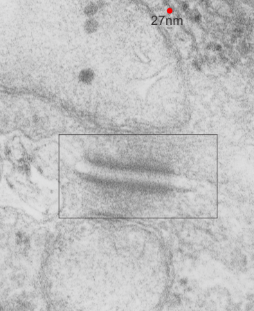
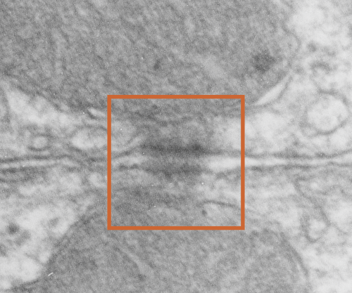
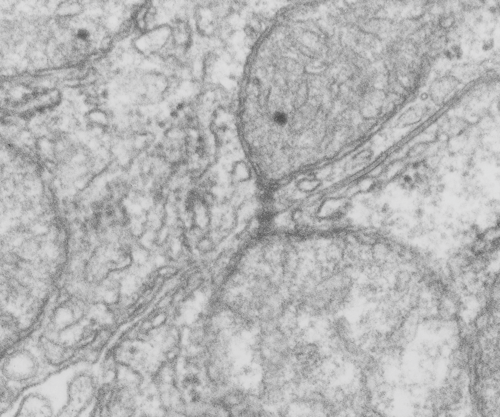
Category Archives: Ultimate order, the cell

The beauty and order of life is astounding.

Rhesus monkey hepatocyte desmosomal mitochondrial tether

There are some ultrastructural characteristics of this desmosome which are interesting. Having a desmosome sectioned opportunely is hit and miss.  This particular section has some structures that look to be the correct size of intermediate filaments that are trailing off the the upper right under the mitochondrion, and there are definite light-dark-and dotted portions of the desmosome (in and including the plasmalemma portion) which have a distinct vertical repeating pattern.

I squeezed the micrograph bottom to top to determine if this was just a shifting of the image during photography or whether these vertical striations would still be visible when i removed (in photoshop) that kind of vertical shift.  The answer was that even then the spacing and the effect of the alternating light and dark areas in the cell membrane portion of the desmosome remained.  Red dot=27nm as determined by ribosome size, and thus the alternating dark and light vertical areas in the section are about 10nm in width.

In addtion, there is a clear darkening of the outer mitochondrial membrane, and a slight darkening of the inner mitochondria membrane at the point of the desmosomal-mitochondrial tethering. One can suggest that there is a relationship between this patterning and the various protein families that comprise the full structure of the desmosome, intermediate filaments and mitochondrial connection.

Baboon hepatocytes and single desmosome

Control animal (neg 5936 block 8830 baboon, f, #23, right lobe of the liver, control bx, modified Karnovsky’s fix, 1% osmium tetroxide, Epon 812, (9 28 1977)). There are a lot of smooth vesicles in this sample and just a little bit of RER, three lipid droplets, and part of a bile canaliculus, possible the space of Disse in the very upper most right part of the image, mitochondria look pretty normal) I was looking for desmosomal-mitochondrial tethers and found this pretty nice desmosome and I pseudocolored the plasmalemma from the two adjacent hepatocytes present at the desmosome purple. There was a clear the “outer dense layer” of the desmosome (which i think is so unfortunately named “outer” and should have been named “inner dense layer” of the desmosomal complex). It had a particularly “loopy” appearance that I highlighted in orange.  Top image is the original micrograph (two hepatocytes) and box is the area enlarged in the figure below  with a desmosome).

Desmosomal-mitochondrial tethers: x2

Here is a gallery sheet of desmosomal mitochondrial tethers, and some are double, some are double-doubles (haha) and some have three tethers.  I can’t overemphasize how interesting these little structures are and how critical they must be for cell function. Just considering how the desmosome itself has become a known mechanism of cell signaling, then the energy provided (mitochondria) and the movement of proteins and mitochondria and signaling proteins via the intermediate filaments, and the latter involved also in cell shape change…. makes these structures very very important.  This gallery (making the gallery itself) caused me great grief… ha ha.. my little desk top computer just didn’t want to handle composite images of over a gig.  Would that I had real equipment…  wonder of wonders.  Enjoy the image(s) of whatever these DES-MiTes  (desmosomal-mitochondrial-tethers)  will ultimately be called (dumber names exist in science).

34 pix, 43 desmosomes, at least 56 mitochondria, numerous species represented here (ferret, rat, guinea pig, stub tail monkey, rhesus monkey, maybe others, and all images are from hepatocyte-hepatocyte junctions)

desmosomal mitochondria tethers and double tethers in hepatocytes


Questions about desmosomes and mitochondrial junctions

Desmosomes:

(who names these proteins — desmosome..  desmo — and soma, that is the easy button, cadherins…  well calcium dependent adhering molecules, i guess thats ok, desmoglobins… not so good, plakophilin, maybe that should be desmoplakoglobofilin.. ha ha…  i guess too that the plaque, and plako…. go together, but there are two intracellular zones (apparently) of the desmosome…  inner (which is already confusing because to me, if the desomosome is the unit being described, then inner would be the closest plaque to the center of the desmosome, which is the central dense line in the intercellular space, but no…someone  named it “backwards” with the center of the CELL rather than the center of the DESMOSOME as the reference point, thus what is named the outer desmosomal plaque is actually “inner” with reference to the desmosome).  It is astounding how differently we all describe our world– and it adds a whole lot of confusion and difficulty to learning and understanding).

More or less facts:
1) electron dense disc like plaques (Stedman’s medical dictionary recommends the spelling “disc” for all medical uses) so I will use disc.
2) extracellular-intra-and-intercellular-cell-to-cell adhesion discs
3) prominent in epithelial cells
4) 0.2 to 0.5 microns in the flat dimension (depending likely on whether the edge of the disc is in the plane of section or the center
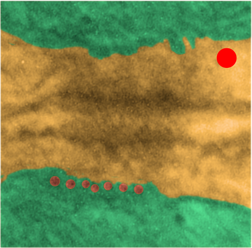
5) very likely different states of cell activity change the dimension and the number and the location of desmosomes as well as the number of mitochondria which are associated.
6) Only a small percent of mitochondria are associated with desmosomes, mainly because there are many mitochondria in each cell. (Volume vs perimeter consideration).

Desmosomal cadherins – desmocolin (3 isoforms) and desmoglein (4 or more isoforms). DSC2 and DSG2 are the most common.

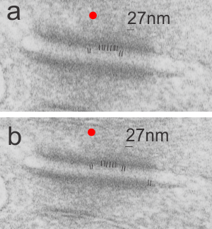
Tying the desmosome to intercellular elements are the “intermediate filaments: a diverse class of flexible filaments that provide mechanical strength to cells. They make up hair, nail, horn, and scale cells, they form the nuclear lamina, which lies just inside the inner nuclear membrane, they span cells to provide strength to epithelial tissues, and they anchor organelles and stabilize the cytoplasm” with these data (Intermediate filaments 6–10 strands per filament (6 was quoted for those which are next to desmosomes), non-polar, outside diameter he quotes as ~10 nm (I think this might be larger, or maybe stains add dimensions)) an edited quote from Methods for modeling cytoskeletal and DNA filaments by Steven Andrews (Physical Biology, Volume 11, Number 1 )

Desmoglein 2

“Desmoglein-2 is a 122.2 kDa protein composed of 1118 amino acids. Desmoglein-2 is a calcium-binding transmembrane glycoprotein component of desmosomes in vertebrate cells. Currently, four desmoglein subfamily members have been identified and all are members of the cadherin cell adhesion molecule superfamily.”

Intermediate filaments – desmosomes – mitochondria

I havn’t read much (or found much to read) about the making – breaking – moving – adjusting of desmosomes, or the energy requirement for desmosome modification, but it seems too much a coincidence that mitochondria would be closely tethered to desmosomes and not provide this function. That said, they have been photographed by me in mouse, rat, neonatal rat, ferret, hamster, syrian hamster, dog, and guinea pig, just to name a few in the list that I have looked at in the last weeks. And they are so similar in all, that so far, if i list one image of a desmosomal-mitochondrial junction from one species I cannot tell it from another. This is good, i guess, but it would be more interesting if the structure showed some variation in structure….. that could perhaps be correlated with differences in the component proteins.

Here is one desmosomal-mitochondrial junction from ferret, which didn’t have a nice even pattern of cadherins in the middle, but did have a punctuated central line…with dots less than 20nm apart…. they seemed a little staggered.  Here is the original micrograph (i removed a scratch in the negative not in the area of the desmosome using photoshop bandaid tool, but it otherwise just a scan of the print. 5940 18648 ferret hepatocyte.

The image below it is an enlargement of the box in top image. Ribosome-red dot for size, and the black lines overlie densities in the intercellular area of the desmosome.


Desmosome-to-mitochondria: connections

More desmosomal mitochondria tethering from hamster hepatocytes. This particular view has two mitochondria, and one shows a line of “dots” or fibers or fibrils in cross section.

This image is from a syrian hamster hepatocyte, and it is just a routine TEM but I noticed (while making my gallery collection of desmosomal-mitochondrial images) that one of the mitochondria showed the repeating dots (cross sections likely of intermediate filaments (maybe types I and II).

Upper image is unretouched micrograph and lower image has been pseudocolored. The latter has green for the two mitochondria that are tethered to the desmosome  at their outer mitochondria membrane (which appears rigid where intermediate filaments are present, and a distinct and definitely separated inner mitochondrial membrane). Along with the intermediate filaments are connections to the desmoplakin molecules the latter being part of the outer dense plaque of the desmosome. The yellow is the cytoplasmic area of the two adjacent cells including the intercellular space and the spot desmosome. Red dot is appoximately the size of a ribosome (27nm) which makes these filaments cut in cross section about 13.5nm in diameter (orange dots in a row) bottom electron micrograph. This hastily made measurement is not that far off from the reported 10nm intermediate filament made up of four monomer filament tetramers (haha… figure that out).

According to Thomason et al,  desomosomal proteins are as follows (slightly rearranged to make more sense to me): Desmosomes have five major component proteins, the DCs (desmosomal cadherins) DSG (desmoglein) and DSC (desmocollin) –DSC and DSG are the desmosomal adhesion molecules. Plakin family cytolinker DP (desmoplakin), and the arm (armadillo) proteins PG (plakoglobin) and PKP (plakophilin).  DP links the desmosomal plaque to the IF (intermediate filament) cytoskeleton, and PG and PKP are adaptor proteins that link between the adhesion molecules and DP.

Who could guess that desmosomes know hebrew?

Was looking at this model for the cadherins of a desmosome and I just happened to see “shin” there in reds and oranges. I love it when nature intersects civilization in an unexpected manner…. so fun, ha ha.  Just out of deference to the authors of this paper on desmosomes, 3D and electron microscopy I will list their reference in case you want to actually see the original. Al-Amoudi, et al., Nature05994 (2007)

More on desmosome shape

“Desmosomes are a complex assembly of protein molecules that form at the cell surface and mediate cell–cell adhesion. Much is known about the composition of desmosomes and there is an established consensus for the location of and interactions between constituent proteins within the assembly. Furthermore, X-ray crystallography has determined atomic structures of isolated domains from several constituent proteins. Nevertheless, there is a lack of understanding about the architecture of the intact assembly and the physical principles behind the adhesive strength of desmosomes therefore remain vague. We have used electron tomography to address this problem. In previous work, we investigated the in situ structure of desmosomes from newborn mouse skin preserved by freeze-substitution and imaged in resin-embedded thin sections. In our present work, we have isolated desmosomes from cow snout and imaged them in the frozen unstained state. Although not definitive, the resulting images provide support for the irregular groupings of cadherin molecules seen previously in mouse skin.” ABSTRACT from Gethin Rh. Owen, Devrim Acehan, K.D. Derr, William J. Rice, David L. Stokes . Some things might be right, others not…. the last idea that cadherin molecules are grouped irregularly i don’t like. (This article i wont read as it is pay-per-view, which really galls me particularly because most research is paid for by tax-dollars and should be free).

” I found a dimension for the width “wide intercellular space (~240 A)” at 24nm and a measurement of the intermediate filament at 8nm (i presume diameter). The 22-24nm (could be less in some instances more like 15nm) as the width of the desmosomal intercellular space (which includes the densities on the outer leaflet of the plasmalemma from both cells and the central dense line of the desmosome.  That measurement looks about right. From this I estimate the width of the electron lucent annulus of the desmosome (which is much more evident when there is a tight junction on either side of the desmosome) to be dependent upon where the section sectioned the spot desmosome, and whether the cut was equatorial or tangent to the spot itself. On one micrograph (stub tail monkey hepatocyte) in which one area of the desmosome is close to a tight junction, shows this lucent area (the outer annulus) to be about 35nm in its radius – that dimension applies to the edge of the central dense line extending to the tight junction. The width of that particular desmosome (where the central line was apparent) was @265nm.  This makes the ratio of the dense desmosomal central line density distance to lucent annulus distance in cross section about 3.5%.

That lucent area may not be a uniform annulus, but if it were even, this assumes pretty much a “round” desmosomal spot weld. In measuring the radius of the annulus (just in the part ring part) I understand that the central dense line of the desmosome will appear in any view with the spot perpendicular to the plane of section, and this doesn’t diminish the fact that closer cuts to the outer edge of the desmosome will have a greater annulus to spot ratio. Fuzzing of the central dense line in the desmosome occurs when the angle of the section is not perpendicular to the spot, and can happen either at an equatorial section or anywere past that equator. So. any question I can ask, someone has an answer to…. so here is the definition of calculating the area of an annulus.. ha ha “An annulus (Latin word for ring) is a two-dimensional region of space bounded by two concentric circles. The area of an annulus is the difference in the areas of the larger circle of radius R, and the smaller circle of radius r. Mean radius (ρ) is the average of the exterior (R) and interior (r) radii. Breadth (δ) is the width of the annulus. “ So I just have to figure out how to use the online calculator.

The ratio of lucent annulus to dense desmosome is not too apparent when there is no tight junction visible right next to it. But that said, I can see a slight increase in plasmalemmal density for a short distance (same type of annulus arrangement as if there were a tight junction there) on each end of the desomosome cross section.