Category Archives: Ultimate order, the cell

The beauty and order of life is astounding.

Perfluorodecyl iodide crystals in vivo: big

Just reviewing some slides of liver of mice given infusions of an emulsion of perfluorodecyl iodide….  some of these crystals which completely fill to overflowing hepatocyte macrophages (multinucleated macrophages or macrophages fused into multinucleated giant cells following the foreign body response to IPFD infusion). Just saying here, some of the IPFD crystals are upwards of 30 microns long, and vary in width from 10nm to many microns. There is a very obvious directionality, always longer than wide. Iin terms of the actual molecular polymerization of IPFD, which is necessary to establish what is “wide” and what is “long” — that remains to be determined. But for now, these macrophages are about 90% “other” with half dozen nuclei in a localized cytoplasmic – crystal free zone together.

And regarding spleen, it has far fewer cells containing IPFD than I would have expected (just a relatively small number of reticuloendothelial cells show collections of crystals (compared to liver from the same animal which does have multinucleated cells full of crystals). So that is kind of interesting.

55 days after 100cc/kg of a 10% IPFD 5%F68 emulsion, the hepatocytes themselves are very well protected from inclusions of IPFD…. there are some probably, but not many. Hepatocytes themselves look nice, but the clusters of macrophages, without eliciting much of a fibrous tissue response are neatly circumscribed.

If…Then

I sometimes get the uneasy feeling that if i see a pattern in an electron micrograph that someone is going to say, ah thats just fixation artifact… and of course they are right. All testing is “fixation artifact” but its the purview of each observer to determine what is background noise and what is real structure.

In the case of looking at perfluorochemicals left behind in tissues, consensus says that there is “nothing” in that empty footprint, just the embedding medium (in this case EPON 812).  So there is a texture to the embedding medium, no question, but nothing I can perceive, and so easily compared to the texture of surrounding tissues, so that when I see a pattern in the tissue I am pretty convinced that it is the presence of the proteins . (They are influenced by the fixative and type and length of dehydration and other stuff not excluding the warping and shifting under the electron microscope.  All those factors play a role). It is one of the reasons that an internal control for size (and i am suggesting background texture) in electron microscopy is really important. I CAN list a short list of variables to be aware of (my knowledge is just observational) which begin even before biopsy, including the time of the last meal, 1) circadian cycle, 2) health of the tissue (in vivo or in vitro –doesn’t matter) 3) mode of analgesia or anesthesia at biopsy (paralytics – solutions with ions (as in perfusion) 4) time till tissue gets into fixative, 5) size of the tissue block, 6) type of fixative, 7) length of time in fixative, 8) length of time and chemicals used for dehydration, 9)type of plastic, 10) section thickness, 11) staining compounds, 12) TEM specs, KV, etc….  so I am willing to accept that not all parameters are under one’s control.

I will however say that the pattern below (in a portion of a macrophage in the liver of a mouse given 10%IPFD in 5%F68, 267 days post infusion) shows a linearity I would not be willing to ignore. low mag micrograph top, for orientation, next below is micrograph of the red box area which shows the periodicity. That is just one of the linear arrangements that are visible in these tissues. rotated image for easier measuring and comparing the textured are (top box) and the basic epon texture (bottom box).

Phagolysosomal and crystal patterns in IPFD inclusions in liver

Two prominent areas of patterning are apparent in phagolysosomes in liver after IPFD infusion. The IPFD crystal itself, and at least four distinct protein patterns in the phagolysosomal proteins. The first image below is an enlargement of the red rectangle, and I have marked off with lines some of the pattern at the long end of the perfluorodecyl iodide crystals which seemed to work out to about 10nm in the short dimension. 23 measurements made at the end are marked off with dotted lines. A red dot=the dimension of a ribosome taken at the same magnification for reference.  Below that is the low magnification image (unretouched — I removed a scratch from the top image but no data were altered as it was not in the crystal itself). Black boxes correspond to four very distinct protein organizational patterns within a single phagolysosomal body which also included the crystal in the top image. I am pretty confident that these patterns are a clue as to which lysosomes are present, and 4 may not be the only ones which show oligomerization into ultrastructural patterns. One thing to think about is that the kupffer cells (likely) in liver acting as macrophages which engulf (and hold onto for months) IPFD crystals will have a different panel of enzymes for lysosomes (and peroxisomes) than hepatocytes, and seemingly can become multinucleated under the conditions of massive IPFD inclusions.

IPFD: phagolysosomes with protein patterned substructure

Phagolysosomes which include IPFD – 1 iodoperfluorodecane aka perfluorodecyl iodide have some interesting lysosomal enzymes.

Of these many enzymes I am thinking that there are many which are “folded” or oligomerized, that is those phagolysosomes which have IPFD show a heterogeneous substructure, zones of tube like enzymes, and areas of mostly untextured protein and then areas of unique coiled ultrastructure.  Below are two electron micrographs, the one on the left unretouched scan of a print, and the one on the right retouched (though I can hardly tell the difference – the one on the right had the coiled nature of the protein burned just a tiny bit with photoshop and several pieces of lint minimized with the bandaid tool in photoshop but no data were changed).  Red dot is ribosome (taken at about 27nm) and the thickness of one of the fibrils is something under 25nm…  which is a little larger than i measured before at 20nm but i am probably responsible for the variation), red bar is 270nm, short red lines are measurements of the diameter of the coiled (polymer or oligomer protein), pale untextured area at the top of the micrograph is the footprint of the IPFD crystal, the whole area is mostly phagolysosome. Inset is the cluster of ribosomes used for size.


Peroxisome in mouse liver after perfluorodecyl iodide

Peroxisomes: I have learned a few basics about these organelles. They are an old organelle evolutionarily, and the enzymes within have become unique to the organisms, species, and cell types wherein they function.  So in the case of the liver of the mouse, there are going to be many enzymes, but basically for the following purposes: 1) for directing peroxiome-relevant proteins produced by the RER into two places, 1) including the peroxisomal limiting membrane, and 2) the peroxisomal matrix.  Matrix enzymes would include those that are protective, in that they metabolize reactive oxygen molecules, and participate in fatty acid metabolism, and peroxisomal matrix those enzymes that have evolved with the species and its environmental pressures and xenobiotics (which would


Inner ear stereo-pair

Many years ago I was interested in reconstructing 3D images from sections of inner ear. I still have many of those slides and images. At that time my son Daniel Miller was into computer graphics. He took my highlighted 2D sections (as shown in the bottom image) and created this stereo pair.  Cochelar duct yellow, ampulla of the semicircular canals white.
It looks like we submitted a cover illustration as well. I cant even remember if it was successful.

Perfluorodecyl iodide: a peroxisome proliferator

Going through archived images of the livers of mice which received infusions of iodo containing perfluorochemicals, I ran onto a hand-written comment which made me laugh. It was “funky print given under pressure to LC Clark (Jr) that perfluorodecyl iodide increases tremendously the number and size of microbodies (aka peroxisomes) in the mouse liver”  (words in italics added today for clarity).  Apparently no one wanted to hear that news in 1973 or 4… perhaps because it was a “damning” mark on the possible use of  perfluorochemical formulations in general, and specifically for artificial – blood substitutes.

Wikipedia summary is here: “They are involved in catabolism of very long chain fatty acids, branched chain fatty acids, D-amino acids, and polyamines, reduction of reactive oxygen species – specifically hydrogen peroxide; – and biosynthesis of plasmalogens, such as ether phospholipids critical for the normal function of mammalian brains and lungs. They also contain approximately 10% of the total activity of two enzymes in the pentose phosphate pathway, which is important for energy metabolism.”
Below are three images, one of liver which shows the very large peroxisomes, in this particular micrograph (not all shown here) they were a volume density of about 30% of the cytoplasm which puts IPFD up there with the best of the proliferators; mitochondria for comparison were about 33% of the cytoplasm. Consensus has peroxisomes at more like 2% of the volume density of the hepatocyte under unstimulated conditions.

The story will be complicated as there are more than 30 enzymes which are in peroxisomes: and the patterning that is seen in the enzymes within the phagolysosomes containing IPFD looks to be similarly wound as the enzymes within peroxisomes in the hepatocytes (top two image groups). From the internet the middle three images (used without permission as a screen print of the insanely egotistical Dr. Jastro TEM collection who most certainly used public funds for this research and collection of micrographs and yet thinks he has ownership of these images…. not civil at all in my opinion and shame on him) is an hepatocyte peroxisome (which i measured against a nearby ribosome and also the micron bar marker, for size) shows a coarse texture, somewhat coiled. The latter and my peroxisome images (top), added from a portion of an hepatocyte from the liver which received IPFD and measured against a nearby ribosome, show similar “oligomerization” or “substructural” protein patterning.

Some protein(s) is creating this texture. It would be interesting to know if the protein in these microbodies (aka peroxisomes) is the same as that which is found in abundance in the phagolysosomes containing IPFD. Bottom micrograph is a portion of hepatocyte cytoplasm from IPFD infused mouse more than 200 days post infusion from which my (top two) images are derived.
electron micrograph hepatocyte peroxisome proliverator perfluorodecyl iodide

Finding the crystal width pattern for perfluorodecyl iodide

I have felt there was a pattern to the width of the perfluorodecyl iodide crystals in the micrographs that I have for some time. Today I decided to measure just two sets of crystals from two different lysosomal structures from two different cells. The graph below shows that there might be a stepwise increase to the thickness of the crystals of something on the order of 18-20nm (or maybe half that). The data from 4-8 measurements along the wide-axis of individual crystals were measured and normalized to the 27nm size of an adjacent ribosome. The mean widths sorted in ascending array and the second plot measured identically (had a few more crystals to measure) is superimposed. The red rectangles might indicate an incremental increase in size. Just thinking on this. I did find a couple of publications on “incremental pattern mining” which would be fun to examine.  Hopefully I will find someone with software already in place to do this. The actual micrographs and positions of measurement are shown below.  Red dots=27nm a presumed size for a mammalian ribosome, in each micrograph. Dont miss the lysosomal protein “snake-like protein with dark central stripe” mentioned in previous which is very clear in the top portion of the top electron micrograph. In this micrograph the values are 18-20 and 9-10 nm respectively for the two-band protein (similarly to previous post HERE).

Endosomes in phagocytes containing perfluorodecyl iodide crystals show unique substructure

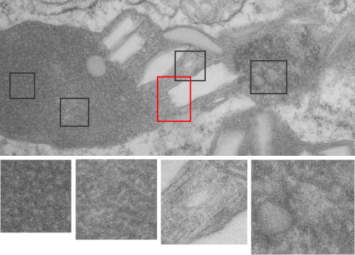
The phagolysosomes in phagocytes which containing perfluorodecyl iodide crystals (in this case the perfluorocarbon has been retained in the body for more than 250 days – mouse liver) show huge amounts of lysosomal enzymes. The collection of macrophages (even multinucleate macrophages) don’t appear to initiate a huge collagen response, and inflammatory cells such as neutrophils, just are not that common, even when phagocytes are plentiful.

The ultrastructural characteristics of the lysosomal enzymes contents within phagolysosomes containing IPFD crystals can vary dramatically within the same cell and in adjacent lysosomes, and within the same lysosome.  This varies from a very fine homogeneous non-textured content in some areas to areas with a more coarse fibril-like texture, appearing like a lumpy (sausage link type) strand or a twisted strand (of two separate elements, or a two strand braid). In some cases the fibrils appear to be solid on cross section, but in other cases more like a tubule. This underscores the variability that would be found in a twisted fibril, allowing for the explanation of both “hollow” and “solid” profiles on cross section.

Transition zones between the types of enzyme ultrastructure are found within the same lysosome suggesting some protein overlap or continuity. There are some areas of very dense accumulation of proteins.

IPFD crystals may be large or small, and most display some obvious directionality, usually the longer side having a thin dense protein (or trilaminar membrane?) coat, but at the ends showing considerable “striping” or dragging out, or whispy periodicity.

IPFD crystals often appear in cross section to be more rounded, or even round.

Some common characteristics of the fibrillar texture of lysosomal contents of IPFD filled lysosomes follows:

  1. relatively even diameter at widest and narrowest areas
  2. often looped and tangled, folded looking
  3. equally even spacing seen as a predominant lucent area surrounding each strand
  4. often running in parallel
  5. higher mag shows sub-layering

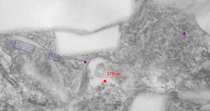
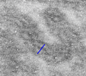
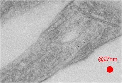
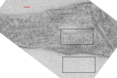
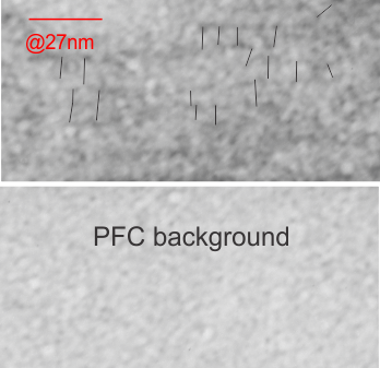
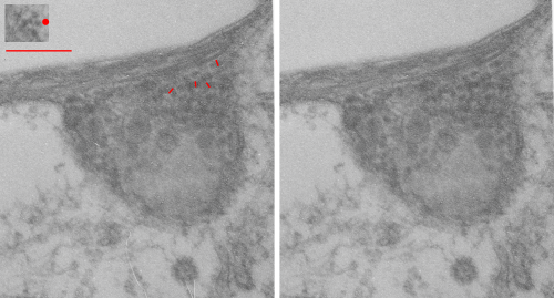
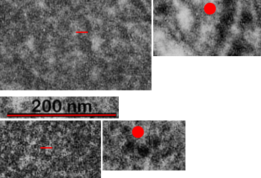
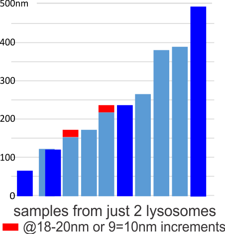
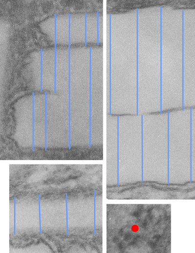
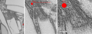
This image below shows a very prominent (yet small in terms of nm) level of periodicity. May not be the smallest since I havn’t be able to find any information on the actual crystal chemical structure of perfluorodecyl iodide), but pretty small for conventional electron microscopy to propose, but this is so obvious that it certainly warrants comment. The single space (inset is same size as lower image and placed on an identical image, enlarged so that the lines (periodicity) could be seen.  The span is about 9-10nm line to line, and the fiber arrangement is double that…. between 19-20nm.  It appears that the protein(s) oligomerized here can come as an interconnected (maybe twisted) double strand.

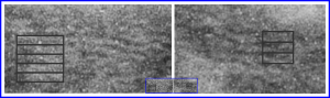
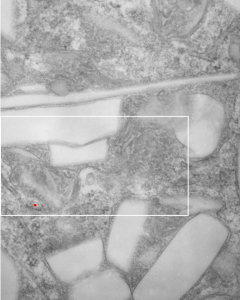
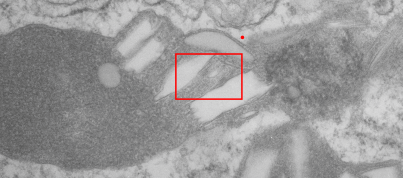
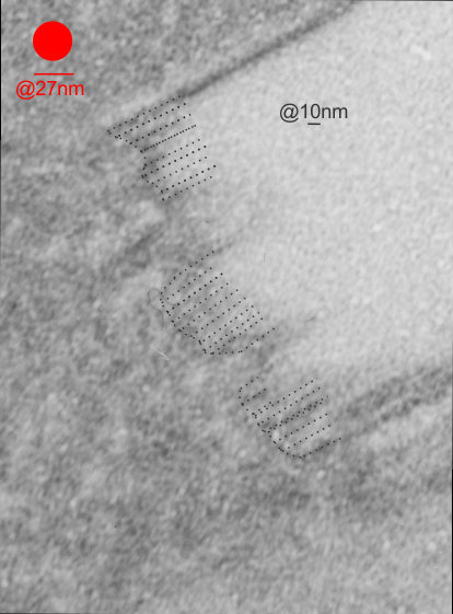
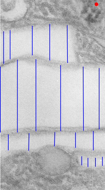
Lower magnification image of the portion of a macrophage from which the inset to the right and above (white box) and the measurements of distance between fibrills (white lines)and smallest periodicity (blue box) measurements were made.  Red dots=ribosomes at an estimated 27nm for reference; blue line = smallest periodicity; purple line = the 9-10nm pair, twisted, representing the fibrills (about 20nm in thickness if section is longitudinal, or diameter ir fibrill is cross sectioned. A measurement of about 20+nm is found from center of one fibrill, perpendicularly, to the center of the adjacent fibrill, (white connected lines on the bottom micrograph show distances between fibrils measured. Image at bottom has blue line of @20+nm –the approximate diameter of the “fibril”, and the dividing line of the two strands is visible.

Perfluorodecyl iodide crystal periodicities

Perfluorodecyl iodide crystal periodicities are something that certainly looks like a phenomenon visible with electron microscopy which sampled such crystals after two hundred plus days residence in mouse macrophages.  The crystals vary in size several orders of magnitude both in width and length, some appearing like little fractures of the whole.On the long ends of the IPFD crystals (within phagolysosomes, and surrounded by lysosomal enzymes which show their own type of periodicity (kind of a tubular or rounded structure — maybe both) protein strands trail out in a very distinct sweeping pattern.  I have measured 15 distances between dense areas in this pattern (marked by black lines in the figure on the right and shown in array as white lines stacked on the left of that same micrograph). Taken back to the mean size of a ribosome (see image on left hand size – red dots are over ribosomes, and equate to 27nm diameter) the space between these strands coming from the long ends of IPFD crystals are about 10nm in width.  I had sort of expected them to be closer to the 15nm measured in previous posts, but this is pretty close, and there is always variability from tangential sectioning and other artifacts.

Speaking to artifacts…. the periodicity here cannot be attributed to some random precipitation of whatever-proteins…. since a clear and even density in background glumping of fixative and lose proteins around during fixation is seen in the “spaces” once occupied by perfluorodecyl iodide.  Comparing with that background…  this is pretty clear patterning.

Left hand image:  IPFD inclusions in a macrophage taken from mouse liver where the animal had received IPFD 267 days prior to euthanasia (details in earlier posts). This is a small portion of a single macrophage and the light areas are the footprints of IPFD crystals. Most of this image, too, is within a single variably-shaped phagolysosome, filled with IPFD crystals and a curley but tubular shaped (oligomerized?) electron dense lysosomal protein(s). Middle images is magnified from box in left hand image, and right hand image is magnified from box in middle image.  Middle and right hand images show the periodicity at IPFD crystal “ends”.   I am trying to find any information on the crystalline structure of IPFD from the chemical databases… but have not found it yet.