Category Archives: Layered intracisternal protein granules in mammalian lung

For 35 years I have thought about this protein organized so nicely in the type II cells of the lung of three mammalian species (guinea pig, ferret, mongrel dog) which I am sure is present in more species, and have not had the time to investigate it further. A peer review publication in the early 1980s was about as far as the study went, though published in an OK journal (Ultrastructure Research) I felt there was more to see than just what I could put down in those few pages. Life makes us make choices, and at this point I choose to play with this old problem. I am not professing anything except diligent “musings” here.

More evidence that SP-A bouquets are found in tangential sections

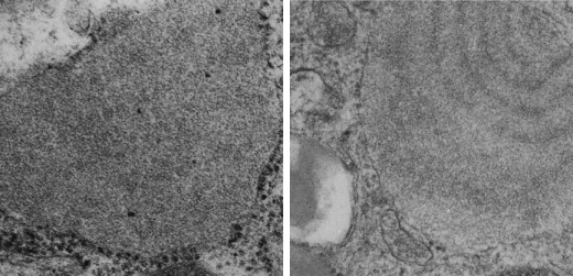
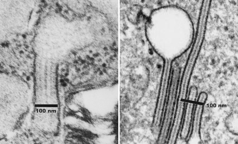
More evidence that SP-A bouquets are found in tangential sections of the protein filled bodies found in some alveolar type II cells.  I am pretty convinced that these bodies are for the greater part surfactant protein A that takes the form of an highly organized (layered) structure that shows linear organization and period repeats that are consistent with an end to end configuration of surfactant protein A, again mirrored.  That is, bouquet end to tail end, tail end to bouquet (times 2) per 100 nm period with the central and outside lines of the periodicity representing the carbohydrate recognition domains.

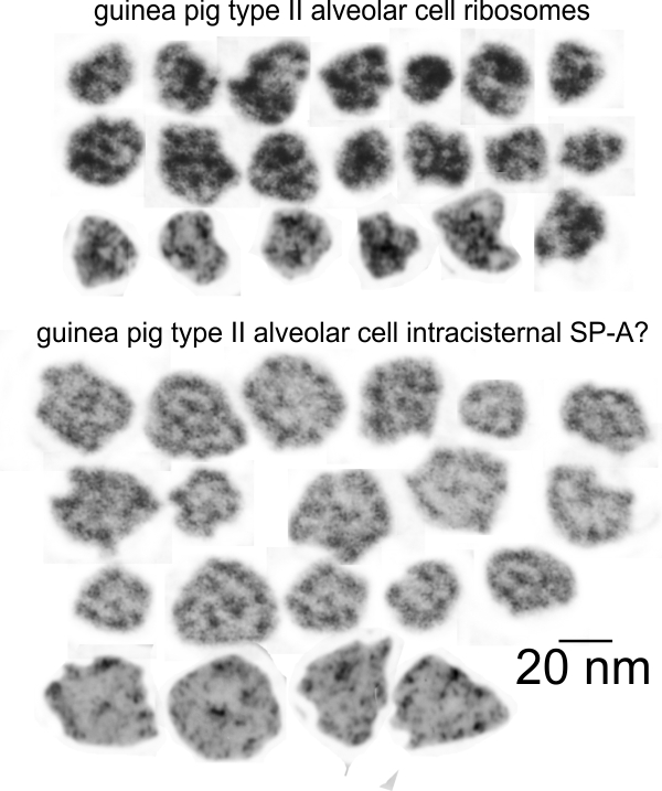
Previous posts show this intracisternal protein on cross section perpendicular to the long axis of the periodicity – or layering, which is easily recognized, and quite orderly, but since the protein inclusions also show curvature and in some cases end to end exchanges in layering direction, and others even as single tubules on cross section, and there are usually  “growing ends” with ribosomes along that growing edge of the RER profile.  The number of ribosomes counted out comes to something on the order of 2 ribosomes per period, and also there are occasions (shown on this blog site) where ribosomes and the protein (presumably surfactant protein A) appear in the same configuration that one would expect from a top down view of the SP-A bouquet right adjacent to each other, it just seems that the anatomy and the protein structures fit together.

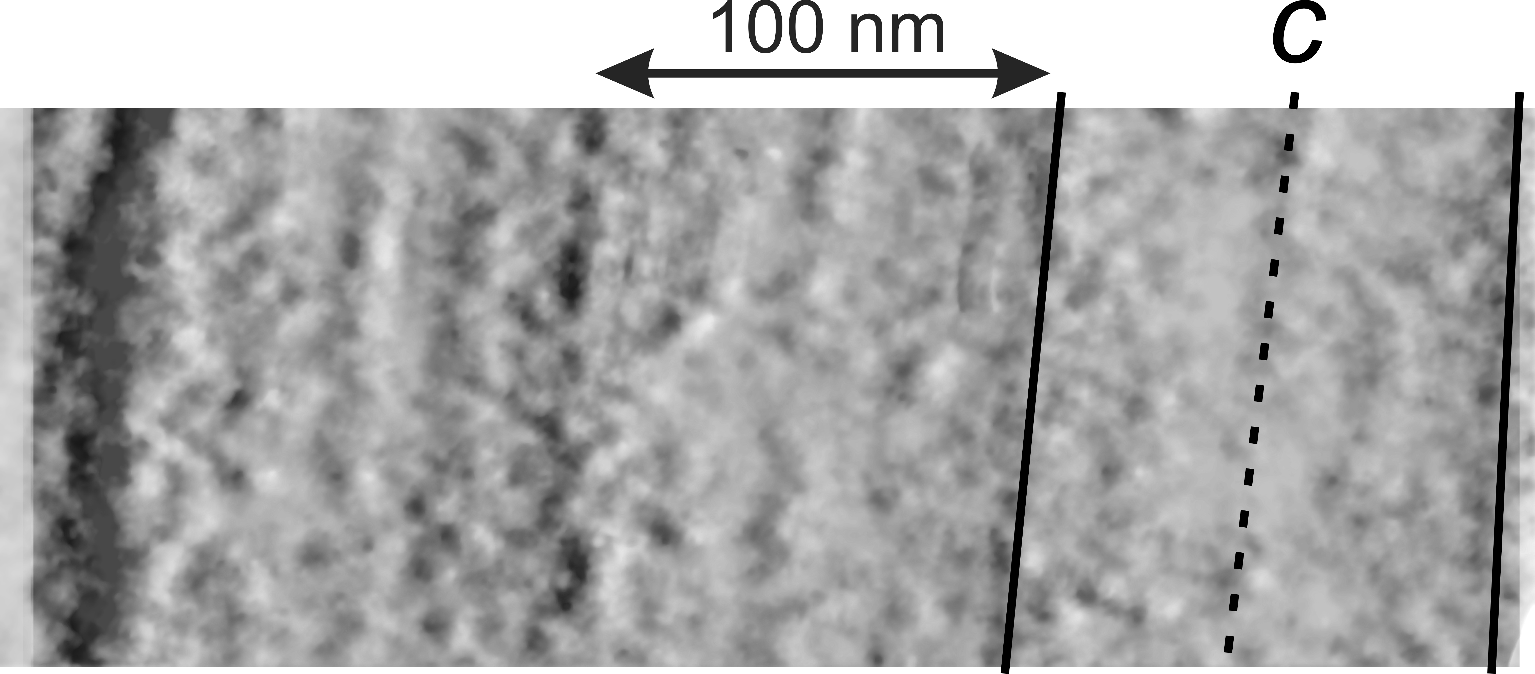
All these new data were found 35 years after the initial discovery of that structure with TEM and that initial search for what this body was, and which started back in 1980 when I queried a respiratory scholar and pediatrician then at CCHMC — who was/is a surfactant specialist — but who also was not interested.  So this study was taken off the back burner in 2016 for a little fun and replay.  I am quite sure that this RER compilation of protein represents an overproduction of surfactant protein A which behaves in the RER profile like other collectins (langerin being a great example and even showing layering patterns onto which a langrin molecule image can be placed, though producing a different orientation of periodicity than SP-A, but conceptually similar, and obviously related in the greater scheme of the way collectins are ordered within the RER.   Here is a quick animation that shows one such tangential section of an RER  profile filled with this protein, but the periodicity  is spread by a tangential orientation to the plane of the section. The darker regions of the image relate to the darker regions of the banding and those areas represent the top clusters of trimers, assembled as the 18 mer of SP-A open and with a center dot presumably representing the neck. Thee micrograph was only contrast enhanced and the burn tool used in photoshop, two scratches were edited out with the bandaid tool, otherwise pixels were unchanged from the original micrograph scanned at 3600 pixels per inch.  Images imported into CorelDRAW, exported as transparent png images, and the animation was made with Swishmax 4. (no data were changed)

Thin sections here are probably about 100 nm thick, maybe a little more, thus there is overlap between one 18 mer and the other.  It has to be determined (but looks plausible) that the hexagonal SP-A stacks up like a honey comb.

Intra-cisternal protein in type II alveolar cells in dogs

Still working on whether this is SP-A (surfactant protein A) in the layered protein paracrystalline type banding seen in the RER of at least three species of mammal. The most prominent of these banded protein products in the RER of alveolar type II cells seems to have occurred in guinea pigs, though they were first observed in ferrets.  These six images are high mag images where just the RER with the layered protein are highlighted (with the black arrow). Since they all came from different images and magnification is just to messy to calculate I have substituted the 20 nm ribosome as a “scale”  so the red dots are a “relative” measurement of 20 nm.  This puts all the intracisternal bodies in perspective.

The dog is different than the other two species in which the intracisternal body in RER of type II alveolar cells is seen as I have only found them with a single banding period…. whereas in guinea pigs the number of periods in a single RER profile with this protein can be very large and difficult to count because of the perpendicular and arched and oft changing directions of the layering.  However, the mean number of bands in guinea pigs, 787 periods,  n=189 RER profiles,  produced a mean of 4.1 (SD=2.9) +- 0.21 (SEM) periods per profile while in ferret of the 625 periods counted, n=123 profiles of RER, the mean number of periods per profile of RER was 2.66  (SD=4.9) +- 0.39.  Mongrel dog had very few profiles, and number of profiles was = to the number of periods (n=7).  Six of those are shown in the figure below:  they were about 100 nm thick and maybe twice that long.

dog_ICBa

More ribosomes and SP-A? in alveolar type II cells

Adding some SP-A profiles (?) and ribosomes from a second tangential section through an intracisternal body.  I am more and more convinced that the major dark band seen in these profiles within the RER of some type II alveolar cells (many of which are posted in this blog) when sectioned tangentially show the bouquet of the 18-mer of SP-A splayed out giving a hexagonal- or oval-like structure.  These are not plentiful, but also not infrequent, and are just prominent enough to be given attention.  As a comparison, I have examined immediately adjacent cytoplasm and do not see this kind of arrangement of the cytoplasm, therefore it seems NOT to be an random artifact of fixation (though we all recognize that fixation induces artifact of necessity) the hexagonal patterns are found in tangentially sectioned areas of the intracisternal bodies.

ribosomes_SP-A-perhaps_more

Ribosomes and SP-A? at RER membrane

This is an interesting picture. It derives from a micrograph from an untreated ferret (my animal # 2, negative 4640, block 18578, 27,900 x magnified 4 x) and scanned and processed in photoshop  using contrast, color balance, dodge and burn only.  While continuing to look for molecules that might be surfactant protein A, the round to hexagonal objects with a central density appeared sometimes to be closely linked with ribosomes at the edge of the RER membrane when the occasional opportune tangential section of an intracisternal body in a type II cell is achieved.

These round SP-A? molecules are more prominent in guinea pig micrographs, but this array along the growing edge of a profile of RER was particularly nice in that several adjacent ribosomes were adjacent to several round-to hexagonal profiles of what might be protein product.

I have highlighted the ribosomes and round structures in blue, put a red arrow pointing the a ribosome, a light red line around one such round to hexagonal structure (left in the figure below) and also shown a blue box around the original cisternal body and ribosomes (photoshopped only with contrast and brightness) (right in the figure below.  The relative sizes: ribosomes, approx 20 nm, the hexagonal structures about twice that.

Most publications give the SP-A molecule as being around 25 microns…. i think if the “bundle of flowers” typically assigned to SP-A 18-mer were to spread out at the head… that the dimension when seen from the top could closely approximate the dimension found here.  Each ribosome can be used as a guide to magnification.

ribosomes_n_spa_at_membrane

Ribosomes and SP-A perhaps?

One transmission electron micrograph gave me a particularly interesting view of the intracisternal protein contents of a type II cell which has the large bodies filled with what I think is an organized layering of surfactant protein A.  This tangential section was unique in that I could find many little objects which comprised a central density and six densities surrounding it…. making it pretty interestingly similar to the “bouquet” style protein 18-mer that is touted to be representative of surfactant protein A.

I have cut and pasted out ribosomes from the membrane of that RER profile for an approximation of the size (which is pretty clearly like other ribosomes at 20 nm in diameter) and kept the sizes relative to these circular hexagonal-with-central density areas in sync.  You can see that they are just slightly larger than the ribosomes themselves, and that the estimated size fot he SP-A molecules (also given at about 20 nm) might be within tolerable limits.

The top rows of the image below are ribosomes and these circular proteins from a single tangentially cut profile of RER (cisternal body) from a guinea pig type II cell;  the bottom set is from a not quite as nice tangential section of ribosomes and the circular proteins from a cisternal body of a ferret type II cell.

Below that image is a low res pair of images (guinea pig on the left ferret on the right) from which the ribosomes and SP-A? were selected.

I included one ribosome adjacent to a round SP-A ? molecule that has its own bar=20 nm.

ribosomes_SP-A-perhapstwo_trangential_profiles

Does surfactant protein A really fit tidily into the four corners of tubular myelin?

Here is another short video as I try to figure out whether the models proposed for surfactant protein A (and D) and some other collectins actually fit what is seen with electron microscopy.  This video is not the best for several reasons of which the main reason is the densities within the tubular organization that are so frequently posted in the literature are not consistent from report to report.  This is to be expected with the different kinds of fixatives, post-fixatives, stains and sectioning procedures, so that is not a surprise. There are sets of images where at least 9 different densities are seen within sets of individual rectangles of tubular myelin (see videos on this site), and some times when only the four corners are dense, and of course there are all variations in between.

The point of this video was to take the actual surfactant protein A, as a proportionally-sized molecule, put it together in the 18-mer and see whether it really does fit (as the flower bundle shown by so many authors) into the four corners of the tubular myelin grid.  The answer was: not great, the main difficulty being that the helical neck region where it connects to the collagen-like portion of the molecule doesn’t really sync up in the real electron micrographs like it does in the diagrams.  (see this video for samples of trying to fit the 18-mer into each of the four corners of sampled tubular myelin grid squares. It just isn’t all that convincing…. but it should be.  I make this one comment.  There is an article which is by Hansen and Holmskov (1998) which actually shadow casts surfactant A and D and shows images.  They comment on the rigid structure of surfactant protein D in shadow cast images, but I am contrasting that with the floppy-ness of surfactant protein A in their shadow cast images.

I would submit that the surfactant D molecules hold their cross like rigor, as depicted in diagrams, where their shadow cast surfactant A certainly does not look like the bouquet of flowers that most diagrams make it out to be.  I am thinking that the floppy arrangement of the shadow cast molecules (pictures of surfactant A which show the 6 bundles of trimers falling randomly all over the place like a loosely held together bunch of balls on strings, is perhaps more reflective of the real situation and that the organization of surfactant A in the corners of tubular myelin is not as tidy as has been deemed.  Video below is produced from composites of rectangles from online images of tubular myelin (in this case i believe the series of images is from a large chunk of tubular myelin within a publication of Hassett Engleman and Kuhn 1980 (without permission).

Surfactant protein A molecules are black and size adjusted to be a relative 20 nm within in one 100 nm rectangle of the tubular myelin. The tubular myelin profiles were randomly selected areas from an electron micrograph from above reference and modified per:  cropping,  rotating, skewing, contrast, brighness, and size.

Surfactant protein A? in this RER cisterna?

This seems to me to be a fit. The relative sizes of the surfactant protein A molecules can be aligned quite easily to the densities, layering and periodicity of the alveolar type II cell inclusions in the RER that I have seen in some of the randomly gathered electron micrographs from studies with guinea pig, ferret and mongrel dog.  Surfactant protein D is almost twice the length of surfactant protein A, at least according to published data and also images from google searches, and would only have a single band in the center for a periodicity.  So surfactant protein D doesn’t fit very well into the banding pattern in the RER of some type II cells.  I can count (depending upon section thickness and orientation) anywhere from three to 9 bands in any period, which fits nicely with four bands of 18-mers of surfactant protein A, accounting for spotty densities of the intermediate lines, and the dark bands where the carbohydrate recognition domains would appear clustered.

This short video takes one of the most detailed image of a profile of RER from a guinea pig type II cell (routinely processed for TEM worked up for a different study entirely)  that I have found in my collection of micrographs,  which is shown in the background, then the fading in is an image of the trilaminar membrane (in good resolution and the relative size is maintained, and the molecular arrangement of surfactant protein A that might be found in these  heterotrimers when they are assembeled into the 18-mer.  (Look at this article for a pretty nice review of the collectins, including SP-A and SP-D molecules –S0REN HANSEN and UFFE HOLMSKOV. Structural Aspects of Collectins and Receptors for Collectins. Immunobiol.,vol.199,pp. 165-189(1998))

Compare: Alveolar type II cell RER inclusion and Birbeck granule

These two images, left is the same alveolar type II cell RER encased cisternal protein that has been made into a short edu-video on this site, and the one on the right is from Grassi et al, J Leuko Biology vol 64, 1998, which shows an intracisternal protein in a Langerhans cell (immune processing cell in the epidermis) called a Birbeck granule.  Some similarities, but many differences exist between these two particular collectin-protein-layered-RER inclusions.  The 100 nm marker shows the big difference in the size of the “between-RER-membrane boundaries for a single period, the one in the type II alveolar cell being more than 2 times wider.  Here is an image, and also a new video.

icb_birbeck


RER protein layering in the type II cell of a guinea pig: image shadowed

The offset shadowing of an electron micrograph (or other photograph) sometimes helps to delineate where the main features are in that photo.  Here the original photo is duplicated, inverted, and 1 pixel by 1 pixel, moved vertically (down) to reveal more detail.  The image is perfectly flat upon beginning, since the images are aligned, and inverse of each other, but the shadow become obvious as the image moves.  This banded protein, which i hope is surfactant protein A or at least mostly SP-A has piqued my interested for 30 years, but I havn’t made/had time to study it.  Here is a video that shows the shadows coming and going, and also marks some of the banding seen in this intracisternal protein.

Shadowed intracisternal body in type II cell

Here is a kind of interesting view of the bands (layering or periodicity) in the intracisternal bodies I am trying to figure out.  This images is created by using a positive and negative image of the identical area, changing the transparency on both to about 80% then taking the negative image (lower layer) and moving it just laterally to create a shadow behind the positive image…. giving the whole thing a dimensional look.  This clearly shows four distinct areas, with a central layer (band), and two identical bands on either side. This speaks perhaps to repeating but mirror arrangement of the molecules along the long axis of the RER membrane. Unfortunately I cannot remember if this is ferret or guinea pig.

shadowed_ICBa