Category Archives: Electron micrographs of liver

Perfluorodecyl iodide crystal periodicities

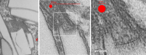
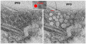
Perfluorodecyl iodide crystal periodicities are something that certainly looks like a phenomenon visible with electron microscopy which sampled such crystals after two hundred plus days residence in mouse macrophages.  The crystals vary in size several orders of magnitude both in width and length, some appearing like little fractures of the whole.On the long ends of the IPFD crystals (within phagolysosomes, and surrounded by lysosomal enzymes which show their own type of periodicity (kind of a tubular or rounded structure — maybe both) protein strands trail out in a very distinct sweeping pattern.  I have measured 15 distances between dense areas in this pattern (marked by black lines in the figure on the right and shown in array as white lines stacked on the left of that same micrograph). Taken back to the mean size of a ribosome (see image on left hand size – red dots are over ribosomes, and equate to 27nm diameter) the space between these strands coming from the long ends of IPFD crystals are about 10nm in width.  I had sort of expected them to be closer to the 15nm measured in previous posts, but this is pretty close, and there is always variability from tangential sectioning and other artifacts.
Speaking to artifacts…. the periodicity here cannot be attributed to some random precipitation of whatever-proteins…. since a clear and even density in background glumping of fixative and lose proteins around during fixation is seen in the “spaces” once occupied by perfluorodecyl iodide.  Comparing with that background…  this is pretty clear patterning.

Left hand image:  IPFD inclusions in a macrophage taken from mouse liver where the animal had received IPFD 267 days prior to euthanasia (details in earlier posts). This is a small portion of a single macrophage and the light areas are the footprints of IPFD crystals. Most of this image, too, is within a single variably-shaped phagolysosome, filled with IPFD crystals and a curley but tubular shaped (oligomerized?) electron dense lysosomal protein(s). Middle images is magnified from box in left hand image, and right hand image is magnified from box in middle image.  Middle and right hand images show the periodicity at IPFD crystal “ends”.   I am trying to find any information on the crystalline structure of IPFD from the chemical databases… but have not found it yet.
Substructure of the lysosomes containing perfluorodecyl iodide

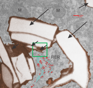
Substructure of the lysosomes containing perfluorodecyl iodide is very interesting. There are issues both with the lysosomal membrane, which in some places may be continuous with other membranes, in particular, i think maybe the RER. Because perfluorocemicals are kind of “slippery” and I think they can travel easily retrograde-style back up into the RER, then it makes sense that some profiles of RER would be seen at sites within the lysosome periphery and the adjacent cytoplasm. I haven’t been able to equivocally find ribosomes on the actual lysosomal membrane to verify that concept.  There are perfluorodecyl iodide tiny crystals…. this is interesting…. an obvious type of fracture or separation plane along the long axis of the larger crystal inclusion certainly is evident….  and i am more convinced that the pattern of lysosomes (a dotted or tubular pattern) is on the order of 20-30nm diameter or width.

Here is an electron micrograph: mouse, neg 9722_10%IPFD, 5%F68 infused 100cc/kg,   8-14-73 sac 5-8-74, 267 days post infusion;  liver. Brown areas=lysosomal enzymes within a bounding membrane, white=IPFD, red dots are ribosomes; pale green is cisterna of RER, green box is area enlarged below.

perfluorodecyl iodide transmission electron micrograph lysosomes

So the bottom two images, left unretouched, right unretouched but with translucent overlays where I see a pattern within the lysosomal enzyme membrane.  Arrows here point to fractured off crystals of IPFD, and the red line is a marker for 27nm which is the same size as the ribosome measured (top inset)…. this makes me wonder if there is some connection between this pattern and lysosomal contents.

Periodicity at ends of IPFD crystals in macrophages

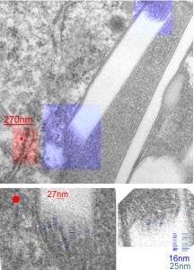
I looks like the lysosomal enzymes at the end of the IPFD crystal inclusion-structures might be tubular. But then as the enzymes pass away from the IPFD itself, there can be a coiling up of the protein with less rigidity.  Measurements at ends of IPFD look smaller than those in the lysosom proper… may be error of just having only measured a single crystal. Same negative and block as previous post, and the ends of the IPFD crystal are colored bluish, and the area measure for ribosome size is in red. Enlargements below, with areas specific measured highlighted. The dense part of the period and the lucent areas beside it.

Texture comparisons: background, cytoplasm and IPFD-lysosome

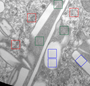
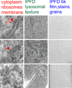
I am trying to determine whether there is a predictable texture to the proteins within phagolysosomes which contain IPFD (perfluorodecyl iodide)(which I am positive there is–so this is academic). I have compared territories from one micrograph with marked inset boxes: red boxes=cytoplasm with a ribosome size marker for comparisons of texture-size); green boxes=portions of the compact and textured lysosomal proteins found just within the limiting membrane of the phagolysosomes which contain IPFD; blue boxes=the background texture of the micrograph found over the “empty” IPFD granules which consists of the acetate film grain and the stains– osmium, uranyl acetate and lead citrate grains. No photographic paper grains are present as this was scanned at 5000 ppi from the negative.

I measured a similar texture pattern from the end-on portion of the lysosomal proteins in last post, and dimensions are similar but not identical… likely my fault, not the fault of the proteins.  So todays measurements are something around the following: dense areas at around 17nm (corresponding to the “spot” in yesterdays post which was around 13nm) and the surrounding more electron lucent space measures about 34nm (which in yesterdays post was closer to 40nm)–all likely resulting from variations in how i measured ribosome size, which is the benchmark.

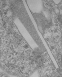
Image below is from a randomly selected phagocyte in the liver (I cannot identify whether this is a Kupffer cell or other phagocytic cell type). These cells can be closely aligned with sinusoids but also near portal tetrads. They are typically “overflowing” with IPFD particles. IPFD stays around for months, unlike other some perfluorochemicals.

Neg 9722 block 3775 10% IPFD 5%F68 injected 8 14 1973 100cc/kg sac 5 8 1974 mouse liver 267 days.

perfluorodecyl iodide electron microscopy 267 days post infusiontexture of lysosomes containing perfluorochemical crystals of perfluorodecyl iodide

Awesome IPFD crystals in lysosomes

Perfluorodecyl iodide was examined once upon a long time ago as a possible PFC for blood substitutes. There are a couple of tissue blocks which I am re-examining to see whether there is a periodicity to the lysosomes that are included in these amazing phago-lysosomal structures, left over footprints as it were, to the presence of IPFD in macrophages (Kupffer cells, fat storing cells?, circulating macrophages, in the liver).

perfluorodecyl iodide transmission electron microscopyperfluorodecyl iodide transmission electron microscopy
This lower micrograph has very definite structure at the ends (one top one bottom) of the crystals. It is a little unfortunate that at the time i printed these negatives (back in the ‘wet darkroom days’ that I didnt use a finer grade paper and developer. Too much grain, but the little indents and rounded areas with a punctate density are going to be relly interesting. I think the lucent areas beside each will measure out the same as some fine lines that I see at the ends of other crystals.

Comparing perfluorocarbon emulsions: Clark et al, 1974

I ran into this publication that I had not even put in my own folder of pdfs, that was published back in 1974, Clark LC, JR, et al, in Microvascular Research. I was sort of surprised to see how much effort went into the assessment of perfluorocarbon blood substitute emulsion particles in the livers of several cats receiving either 1) no emulsion, 2) PP5 emulsion or, 3) FC47.  Early on it was evident to me that the re-emulsification of fluorocarbons in vivo was quite dependent upon the chemical characteristics (but i dont know which) of the PFC, combined with — or prompting the lysosomal response, coupled with the apparent diffusion and aging characteristics of particles within phagolysosomes.

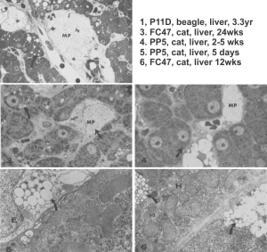
I have extracted the micrographs from this publication and added the descriptions (which were unfortunately difficult to find) from the publication which shows that P11D and FC47 clearly differ from PP5 in the responses they elicit from the phagocytes in the liver (likely Kupffer cells and fat-storing cells, are RE cells. Top three images are light micrographs, bottom two images are transmission electron micrographs.  No bar markers unfortunately but in the top three use a nuclear profile as about 15 microns, in the bottom two, use the short diameter of a mitochondrion as about 0.2 microns.

perfluorocarbon emulsions liver transmission electron microscopy PP5 P11D FC47In this publication Clark notes that PFC ring compounds with O, and N, and straight chain PFC with O, hang around for a more extended time than those with just C and F.

CAS names for the iodine containing perfluorochemicals investigated for use as artificial blood

CAS names for the perfluorochemicals investigated for use as artificial blood (blood substitutes) are really important, as so many acronyms appear for the same chemicals. I have to say it has been a major headache just going through notes, and posts, and publications, to figure out the long names and CAS numbers for the acronyms used in the 1970s.  I do thank the kind person at Fluorox labs (he might not want his name mentioned? i did not ask him) for some valuable info, and of course being a chemist, he suggested that I only use the CAS names (which i will try to do).

Two perfluorocarbons with at least one iodine atom, were made into emulsions and infused into mice in the 1973-1974 range by Leland Clark Jr (and the research faculty and support in his laboratory) one was called n-PF alkyl ethyl iodide (PFEI for short) and Fluorox Labs gives CAS 68188-12-5 to this compound and a second compound Perfluorodecyl iodide (IPFD) – presumably 1-iodoperfluorodecane) has the number CAS 423 621.  I am really curious to see how these differe in vivo, since at the time, no one seemed to “have the time” to look over differences.  There were 4 mice total, one mouse gave up several different tissues, from the others, only liver. Details below.

n-perfluoroalkyl ethyl iodide (PFEI) (CAS 68188-12-5)
Blocks 4759-4762 mouse 4, liver (one list says 4759,4760), liver
emulsion# 740313; PFEI 10% F68 5%, infused at 50cc/kg,
infused 3-13-1974 –sac date 1-14-75 (307 days)

perfluoro decyl iodide (IPFD) (CAS 423 621)
emulsion #730814, log#86 IPFD 10% F68 5%, 100cc/kg
2339-2342 mouse 1, liver (no osmium)
Blocks 2343-2346 mouse 1, liver
Blocks 2347-2350 mouse 1, kidney
Blocks 2351-2354 mouse 1, spleen
Blocks 2355-2358 mouse 2, liver (no osmium)
Blocks 2359-2364 mouse 2, liver
emulsion #730814, log#86 IPFD 10% F68 5% 10cc/kg
infused 8-14-73 –sac date 5-8-1974
Blocks 3774-3780 mouse 3, liver

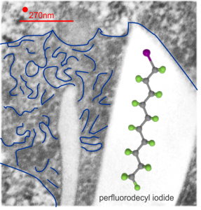
This is what I believe is the structure (inset within the large, presumed, crystalline footprint formed by IPFD; grey carbon atoms, green fluorine atoms and a fuchsia atom being iodine). Thanks to Fluoryx labs and Chemspider and wikipedia and of course google.

Two white rhomboid spaces where IPFD was once in these lysosomes are seen (vertical spaces–one large one quite thin), and the blue outline is the apparent limiting membrane of the lysosome (which encloses the crystalline IPFD), and the blue lines that wiggle within the lysosome show a sheet-like-periodicity that needs further investigation but is very clearly a pattern in most of the lysosomes with IPFD inclusions, and not really present in lysosomes from the other artificial blood substitutes that I have looked at with TEM (that would be a long list).

Red dot is ribosome to estimate size at 27nm, red line is micron marker derived from ribosome size. No measurements yet made on the longest crystal length, but it is going to be many microns for the largest, and the directionality has yet to be associated with any cytoplasmic elements.

electron microscopy perfluorodecyl iodide

Perfluorodecyliodide in a lysosome

This is an interesting perfluorochemical (IPFD) and in the mid 1970s it was used as an emulsion for studies on artificial blood (blood substitutes). It was the only perfluorochemical used that had a crystalline structure in vivo, and while i have posted previously on it, i have hunted up about 10 negatives (i think the only negatives i have) from those long forgotten files and will examine whether or not there is any peculiarity about the enzymes, perhaps even a periodicity to the lysosomal enzymes which surround the IPFD inclusions.  I have to assume that the cell in which the crystals are appearing is either a kupffer cell or an hepatocyte, I can’t be sure which.

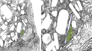
Just from the beginning, here is an image scanned at low ppi, but it shows a curly look to the lysosomal structure surrounding this narrow IPFD inclusion.  IPFD is purple outline, green is the lysosomal contents. Image on right is enlarged from left.

perfluorodecyliodide in lysosome

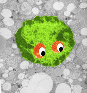
Little green aliens among us

So sorry, ha ha, but when i see this kind of nuclear symmetry (an electron micrograph, with two little presumptive eyeballs staring at me (condensed chromatin), zygomatic arches coming off (as condensed chromatin on the sides of the nucleus), and a hairline part at the center of the frontal bone (condensed chromatin and nuclear pores on the inner nuclear membrane at the top of the nucleus) and a pointy chin, I am compelled to laugh out loud and add this to my collection of TEM-Devils, and sometimes other objects like hearts and ghosts and snowflakes. Anyway, here is a little green cutie, which spawns from a mouse hepatocyte null for 14CoS and not receiving NTBC as a “saving” medicine and prone therefore to undergo massive hepatocytic apoptosis within a day or so of birth, and ultimate demise. This mouse is 24 hours post partum and showing signs of hepatocyte apoptosis, and mitochondrial and ER disarray, neg 16042, block 65718, anm#5, one chatter line removed with photoshop, layers constructed in photoshop, pseudocolor and eyes added in CorelDRAW.  Dont you wish there was a place to publish such stuff…. Maybe i will start my own journal…  help me think of a name.  (LOL)


Outer nuclear pore filaments and mitochondrial proximity

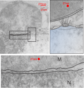
These little junctions, that is, nuclear pore – mitochondrial associations, have been fascinating to me.  I have noticed and photographed (whether ideally fixed, or in focus, or in this species or that cell type, just as many as i can) to try someday to figure out what tasks they are carrying out. The obvious of course, providing energy, maybe for nucleo-cytoplasmic transport, but maybe too for nuclear rotation, as we all know happens from observing those old time lapse videos of tissue culture cells with mitochondria dancing close to the nucleus and the nucleus spinning  — ha ha – new question, to nuclei on the northern hemisphere rotate the same way as nuclei cultured in the southern hemisphere?) and also for nuclear shaping, but importantly, do they have something to do with chromosome territories? That is, something more than just an indirect influence like shape or transporting molecules.  Might they participate in the transport of ions through the outer pore areas as well as the core.  Might they influence assembly and repositioning of nuclear pores, making and or breaking them.

This particular nuclear pore-mitochondrial association is not the best micrograph in the world by a long shot, but it is from a heterozygous animal conditional ko of  Gclc in the liver (wc/ii) so has some increase in oxidative stress.

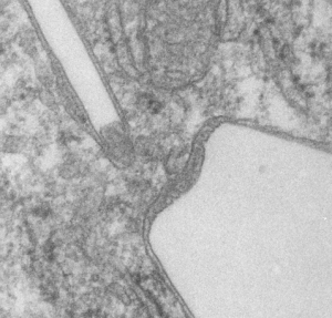
The original micrograph is on the left taken at 9500x, neg 17535 block 73458 anm# 505 wt hepatocyte.  filaments (wiggly lines) going up toward the mitochondria (top shaded grey) are prominent , the nuclear basket is not prominent, and something looks to be a pattern underneath the pore (black oval) that would likely be some tangential portion of the nuclear membrane (not hazarding a guess as to whether outer or inner.