Category Archives: Desmosomal mitochondrial associations

Desmosomal mitochondrial tethers

It makes a lot of sense that there are mirror images between two cells  — that is in terms of desmosomal / mitochondrial tethering. I have seen as many as six desmosomes tethered to two mitochondria (in juxtaposed cells). It seems that if the mitochondria supply the energy and the Ca+ regulation (for unzippering the desmocollin and desmoglein) and signals for dissolution and/or construction of desomsomes that one mitochondrion could do the job for three or more desmosomes.  I can envision the mitochondria alligned up along the lateral plasmalemma like soldiers standing at attention ready to build or destroy…

    1. seems pretty certain that transmembrane  portions of desmocollin and desmogleins affect the trilaminar architecture of the lateral plasmalemma, clearly seen on electron micrographs — they stiffen it….and widen it.
    2. There is an also an effect on the lateral plasmalemma in the annulus of the desmosome, appearing to be slightly more dense than plasmalemma further away from the desmosome, and also with slightly wider extracellular space (than non adhering areas) from outer lamina of the plasmalemma of each adjacent cells.
    3. Seems pretty likely too that most of the diagrams i have found in publications on the topic which bring intermediate filaments to the desmoplakin molecules at a parallel or nearly parallel position are just not accurate. I see lots and lots of perpendicular structures, that is desmoplakin perpendicular to IFs, rarely if ever one that looks like the desmoplakin and IFs are running the same direction. In fact, it doesn’t make sense architectural sense (in terms of adding resistance to pull and shear and separation to have them run the same direction. The long polymers (the IF) run parallel to the mitochondrion which really remains a fixed (seemingly) distance from the outer desmosomal plaque proteins…and plasmalemma. It looks likely that there are 4-6 IFs or more that run parallel to the plasmalemma (and outer membrane of the mitochondria)  not perpendicularly to it.  Just my thoughts (and what I see).
    4. Here is an electron micrograph which has cute little cross sections of IFs in the space between the mitochondria and the desmoplakin and outer desmosomal plaque proteins. (see the blue dots in micrograph on right, which I interpret to be IFs). Left hand image unretouched from scanned photo. (I could have added at least 5 more blue dots to the image on the right where blue dots are overlying cross sections of IFs.)

Intercellular organization of desomosomal proteins

Looking further at the great publication by He, Cowan and Stokes in Science 2003, i just was surprised to see what looked like the most orderly set of dots in the intercellular space on either side of the central dense line (where the desmocollins and desmogleins are supposed to be hooking up in their “velcro-like” attachments. I did a screen print of their micrograph (top image which I did not manipulate)and pasted into photoshop to enhance the pattern with the burn tool (shown in the two bottom images) as an overlay of two separate layers of their original image.  The dots and grid are so obvious as to be almost “silly” and if you don’t see what i am talking about…. check out the red dots within the grids in the two images below their micrograph.  I have looked at hundreds of desmosomes…. now i have to go back and see whether any of my more opportune images show dots within the grid (which I have seen before.  Here the Y and alternating pattern of the intercellular grid is very very apparent.

It is still possible i think, to have the Y formation be the most prominent, and also repeated in a very orderly fashion (one difference between what i see and what the paper above suggests… when they call the desomosomal cadherins to be in “a knot”.  I don’t think disorder is part of this structure… but there rather there is an order which is just a little difficult to detect, owing to the very many angles (at a thickness of 90nm) to bisect a desmosomal spot and untying those possibilities (not to make a bad pun on their title) is not easy.  U havn’t yet found a publication that speaks to anything that would make these central densities.

I know there are no mitochondria in their micrgraphs… and that is ultimately what I am curious about, but just in case you havn’t figured it out yet…..  biology is totally amazing.

3D intercellular space of desmosomes

This is an awesome representation of the intercellular space of a desmosome (that I am not giving a classification to…. either coming, stable, or going since apparently the intercellular area undergoes ultrastructurally visible changes, namely the loss of the central dense periodicity, as they change states).  The paper by He, Cowin, Stokes appears in Science linked here. Two reasons that I love but dont like this diagram. 1) it depicts exactly what can be seen with traditional transmission electron microscopy of the intercellular area of desmosomes, but 2) it doesn’t seem to ring true that the organization since a “knot” is less orderly? in my mind, more “chaotic”, and I think the electron micrographs show the intercellular organization is way more orderly than the diagram suggests. 3) the number of molecules in the colored diagram (part A as they label it) is more than I seem to see in the sections with TEM.  The banding (i am guessing most agree to be desmocollins and desmogleins) is so regular, and the space between so lucent, that all the knot like other molecules are likely overrepresented.

I agree that in this particular view here there is only a tiny hint of any symmetry. I do like that the dimers that they have highlighted (one actually as a lambda, my favorite representation, and the one that fits the intercellular space-repeating order of a desmosome in many of my own electron micrographs best —  lambda shapes mirrored vertically and ofset by half). There is no way here to tell if something was influencing the desmosmal structure (e.g. the presence of a very close mitochondrion, maybe participating in the formation or destruction of the desmosome in their picture).  Lots of opportunity for that variability to be a part of a process influencing what is viewed as a chaotic knot which in fact was a highly organized intercellular component of the desmosome. I am linking their image to their article.

(it would indeed be fun to have access to that equipment, and knowledge of how to use it, and possess the specimens made to order,  poof, all at once and with extreme precision).

This publication also shows some TEM images of desmosomes which i did some measurements with: the distances between the densities at the outer plasmalemma of both cells, and the distance between the periodicities of the central dense line of the intercellular space. All distances were similar… mean of all was about 4nm center of density to center of density.  The intercellular width (from the outer plasmalemma to the adjacent cell was about 9nm. See lines from that TEM below (link to the manuscript is above).


How does a cell know its “equator”

Here is a great photo from Nature Reviews Molecular Biology.  I was looking at the intermediate filaments (red) and understanding the effect of looking through 3D space, with fewer filaments on either side of the central area where the nucleus resides.  There is no equivalent effect seen with the desmosomes… they only occur at the widest part of the cell….So tell me… what sense (other than the tension and arrangement of the intermediate filaments (which also are pointedly connected to the nuclear pores) do they “feel” that tells them the widest part of the cell.  In stratified epithelia, there are many belts… .but this is tissue culture… only one belt.

Without question, cells in culture can sense “confluence”, but this is still a single layer, and the belt is the equator (but the shape is not spherical (haha).  Awesome. Some have suggested that confluence is required for mature desomosomal appearance.

Intermediate filaments: the great connectors between and within

I dont know exactly how to go about diagramming this…. but it seems that the filaments (no surprise should be had) are so critical….  for so much in cell function, just in orchestrating all the comings and goings of the organelles, in addition to some filaments making sure what enters the nucleus through those cute little nuclear pores and regulating the presence of mitochondria (energy, calcium, apoptotic proteins and membrane potential) at nuclear pores and at desmosomes (maybe adherens junctions as well).  All in all, a highly complicated and interesting topic.

here are a couple of better diagrams i found on google


Early article on desmosomal-mitochondrial complexes

After so many years of looking for articles on this topic (I actually wondered how this phenomenon could go unnoticed for so many decades) i did find one manuscript (which references two others) by Deane et al 1966 which mentions them and shows several micrographs (sometimes pretty low manification). They found these first in fetal and neonatal tissues, so this is a clue and also important for the concept that I have which is that in tissues with higher rates of cell division that also are epithelia which connect at environmental interfaces (meaning the “outside” whether that is physically outside or not) have an active mechanism for building and disassembling desmosomes.  They felt it was a one-way passage…. i think it is more a modeling-remodeling phenomenon.  So this was nice but bitter sweet at the same time since I thought my early paper in a very obscure journal in the 1980s was the first.

(I am going to use this acronym…desmosomal mitochondrial tethers  regardless of how silly it sounds just because it is too time consuming to type out 32 characters when 7 will do, my apologies.)

miller et al, desmosomal mitochodnrial interactions_1985
deane_1966_desmosomal mitochondrial complexes
In figure 1, and enlargement inset called figure 2 (a portion of which is below) of their article, they make a cute comment…. about a desmosome beginning to develop nearby to a reasonably well defined desmite. It would be better perhaps to have recognized that there were two possibilities — one, that it was a tangential cut to a sequentially placed tether along a single mitochondrial outer membrane, and the other that it was a desmite “being built or broken”. This little area of not completely well defined junction they label with a black arrow. You can see that it is not quite a desmosome and the mitochondrion really isn’t that close to it.  The label TF (i don’t know why it is in italics) surely stands for tonofilaments…. which I refer to as intermediate filaments.


Just using their image and their micron marker (0.5mu = 500nm) it seems that their cut in this desmosome is just off center but close (diameter with the annulus) at about 286nm (central cut yields a desmosome of 300nm) and a diameter of 214nm excluding the annulus.  About 162nm span the distance from the outer mitochondrial membrane to the plasmalemma membrane of the desmosome of the cell on the same side. A quick measurement for the thickness of the extracellular space of the desmosome in this image was about 18nm.  About 15nm /2 for the dimension of the annulus area on either side of the desmosomal spot. This micrograph shows unequal annulus dimensions.  Lovingly I point out the scratch (likely on the old acetate negative) lower left side of the desmosome… and in sympathy say, my negatives and micrographs have many such scratches. One thing to not is that 3-5 intermediate filaments are lying almost parallel to the plasmalemma and the outer mitochondrial membrane… certainly NOT like the diagrams seen routinely  — see my post with desmosomal diagrams with hairpin (wrongly directed) lines for intermediate filament attachments to desmoplakin molecules.

Desmosomal symmetry: not a complete mirror images side to side

Desmosomal symmetry: not a complete mirror images side to side, since it seems to occur in my micrographs, and obviously too in this micrograph from Green and Gaudry, that the separate cells (adjacent cells bound by the single desmosome) may have a propensity to have intermediate filaments coursing by at perpendicular angles.  Maybe also chance – I guess it is a 50 50 chance to be chance (haha).

But also in this quote from Green and Gaudry.…I take exception to their remark that the desmosome is tripartite. A quote from their paper yielded this text of which several parts make no sense to me “electron micrograph further illustrates the highly organized ultrastructure of a desmosome (yes, i agree) in which mirror-image (partly agree, but not always true), tripartite electron-dense plaques (show me 3, ha ha, and are you counting lucent bands, plasmalemma (not really to be counted and intermediate filaments?) sandwich a central core consisting of adjacent plasma membranes (wait, plasmalemmas are NOT part of the central CORE, and what is meant by CORE anyway, that is a term that needs definition) bisected by an intercellular zipper-like midline (yes, zipper like dense midline -i agree)

They didn’t take into acount the different directions (which may be purposeful) of the intermediate filaments coursing by the desmoplakin molecules in the adjacent cells… as sometimes they appear as 10 nm cross sections and other times as low arcing swooshes. I ask, what part of the plaques are divided into 3??? This is a confusing, since there is nothing distinct about the 3 parts they refer to… they could represent many/or any different layers of this organized structure. Their own electron micrograph (pasted below) shows pretty convincingly that there is not total mirror symmetry to the desmosome since their cell on the bottom part of the image shows cross sections of intermediate filaments while the cell on the top part of their image shows the longitudinal swoosh of intermediate filaments.

One thing that their electron micrograph shows that is rarely commented on is the desmosome annulus… this ring which of plasmalemma which is just slightly morphologically different than plasmalemma further from the desmosome.

While on the topic of their diagram, i think more care could have been extended to the depiction of the tight junction which is really a weld, and they show it as a wider structure than the two blended plasmalemmal membranes really are. And the adherens junctions are diagrammed to be as prominent as desmosomes, which they are not, and the labeling of adherens junctions actually appears closer to their green blob desmosome and thus doesn’t really direct attention to the junction they are wanting to show.  Maybe that works for a visual aid for some, but for me it does the opposite…. working from a place of knowing the actual structures and trying to figure out what visual errors the “diagrammer” has created… is frustrating.  This brings up the legion of diagrams of really poorly drawn desmosomal structures… ha ha.  I do wish more care could be taken in scientific illustrations..N
Not a single diagram that I have seen on desmosomes and intermediate filaments has mentioned the importance of the presence of mitochondria in building and breaking down these structures.

Desmosomes: ductile, double-sided, shear load rivets

Desmosomes are kind of like Oscar rivets, and are blind perhaps, or double-sided. the latter have splits (the desmogleins and desmocollins) along the shafts. These splits cause act like flares and join in the central dense line (periodicitities) of the desmosome. The plakoglobins and plakophilins form the “head” of the rivet along with desmoplakin, and these rivets are unlike most rivets found for metal and wood work, because they are flexible, and have a  “spring-hinge” at their head, the desmoplakin-intermediate filaments junction (which i believe is perpendicular not parallel (as most of the diagrams show), which can flex and bend and allow for motion of the desmosome as it gathers together as weld between two cells.

Below is a set of diagrams taken freely from the internet from publications on desmosomes (including my own). It can be seen that the majority of diagrams don’t really “see” what the microscopist sees as the course that intermediate filaments take when they are adjacent to desmoplakin.  The diagrams on the lower left, and all those in the center and the lower right have intermediate filaments coming from the cytoplasm and making hairpin turns at the desmoplakin molecules…. However, the micrograph on the upper left, and the diagram in the upper right have the intermediate filaments which actually appear to arc ever so slightly, to be NOT forming hairpin turns to desmoplakin, but gently curving perpendicularly to the desmoplakins.

It seems more likely that the type of shear stress (the connection between desmoplakin and the intermediate filaments perpendicular to one another) would be more advantageous at binding two cells together than the diagrams below (with hairpin turns) which suggest that the connections between intermediate filaments and desmoplakin are parallel, thus involved in tension stress.  The former intuitively suggests more efficient shock absorption over a smaller distance than the latter, and actually looks more like the actual anatomy than the latter.

The disparities among the images from electron microscopy and the diagrams from the literature are the reason that I began to try to think this through. Intermediate filaments are seen in micrographs that look perpendicular to desmoplakin (like what is seen in the micrograph in the upper left part of the diagram as brown lines), or as 10nm round dots (if they are cross sectioned).  Any ‘apparent’ long intermediate filaments parallel to desmoplakin (as seen in the bottom center electron micrograph) can almost always be explained as tangential cuts.

This arrangement is most clearly demonstrated when intermediate filaments form a flat perpendicular band between the desmoplakin molecules of the desmosome and the mitochondrion when the desmosome and the mitochondrion are tethered together.
diagrams of desmosomes

Desmosome dimensions relative to adjacent membranes and mitochondria

It seems to me that there is a pattern of thickness  changes(width or height if you wish… because of the orientation of the diagrams below) and the rigidity of the plasmalemma inherent in the desmosome (likely due to the transmembrane parts of desmoglein and desmocollin ) and that rigidity includes a specific intercellular space dimension. I have seen this published at about 34-38 nm (that is, from the inner leaflet of the plasmalemma of one cell to the inner leaflet of the plasmalemma of the adjacent cell) to be something on the order of 38 nm.  Using that dimension, if i measure from inside plasmalemma of one cell to the adjacent cell and compare that to the width from the same places in the 200 nm ring or annulus around the desmosome there is going to be a change in the dimensions (which i could measure as just a few nm greater than that within the area of the desmosome proper. And, then a reduction in the intercellular width (per the more routine proximities of two cells) which becomes something like 40 nm.

Here is an article which suggests some dimensions for the desmosome, but does not address adjacent variations in the plasmalemmae including the anulus.  You can compare with two images and measurements below. Keep in mind that the two images below are not derived completely randomly, as I picked two which had substructure which was clear enough to measure.  The annulus of these two desmosomes is indistinct, and longer than that seen in other desmosomes. This likely relates to the plane of section on a perpendicular axis of a round desmosome and surrounding annulus (the latter being increasingly seen as one sections nearer the periphery of the actual desmosome).

top diagram shows a length of two adjacent plasmalemma, measurements relative to that image, bottom one is a different desmosome, notice brown lines for intermediate filaments, black line for length of the desmosomal spot, dotted lines at periodicities of the bridges between desmocollins and desmogleins, outer dense line with dots, periodicities of the extracellular membrane anchor, pink bracket, thickness of the desmosomal intracellular elements, which includes proteins of the intracellular anchor (desmocollins and desmogleins) plakoglobin, plakophilin, desmoplakin and intermediate filaments (visible at the desmosomal-mitochondrial tether on the bottom more clearly than the desmosomal-mitochondrial tether seen at the top of the lower micrograph.  While a big deal is made of the inner and outer dense plaques of the intracellular part of the desmosome, the lower portion of the lower micrograph doesn’t make that case.  Were the micrograph sectioned end on to the intermediate filaments below, there might be a more visible inner dense plaque.  The outer dense plaque (plakophilins, plakoglobin portion and desmoplakin proteins) is well defined. NB, there is a “flatness” or “rigidity to the outer mitochondrial membrane where the intermediate filaments lie beside it… i hope to search for proteins that might be involved in the linking of mitochondria and intermediate filaments.


Desmosome dimensions

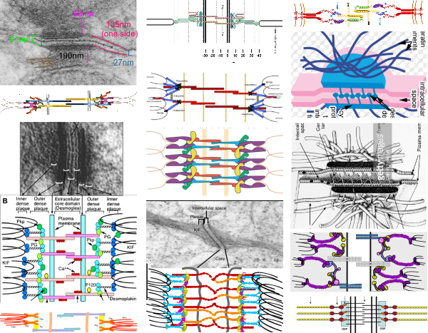
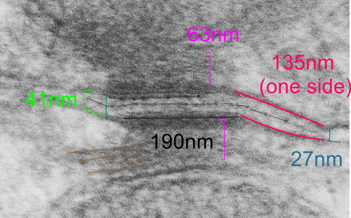
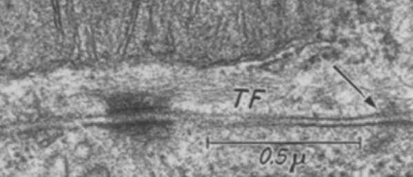
I found a cryo-EM paper of desmosomes that was pretty nice.  Well desmosomes in the lower portion of the epidemis might be like desmosomes in liver, and it is very clear from this paper and others that the desmosome is adaptable, become different things in different cell types and tissue types. So this is a general description, but the basics are present in desmosomes of the liver.  The measurements they show do NOT exactly fit what is stated for the “viable epidermis  desmosome this publication states, but they are pretty close to those that I have re-measured just from their image, their bar marker, and my own repeated measurements from densities to densities.

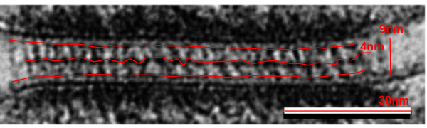
The measurements I came up with are posted on the micrograph. The baseline measure came from the micron marker given for their own image. The measurements I made are for inner lamina to inner lamina of the two plasmalemmas; outer lamina to outer lamina for the two plasmalemmas (extracellular width); the pretty lucent area just extracellularly to each plasmalemma, the length and dimension between the desmoglein and desmocollin molecules (cant tell which is which) and also the “v” type structure, alternating and overlapping as the center dense line of the desmosome (in the extracellular space).

I thought it was interesting that one researcher called this intercellular linear pattern random, while another called the pattern linear…. in fact i think they both missed the pattern and I have outlined the cadherin rhythm in green. The distance between the repeat portions (ectodomains) of the latter looks maybe to be close to linear, or slightly curved, and the N terminals (to my way of thinking) create a picket fence, or zipper kind of pattern.

The micrograph I used for these measurements came from a paper by Ashraf Al-Amoudi, Jacques Dubochet, and Lars Norlen which is available online. Their bar marker is at the top (50nm) and all other measurements were made from this distance.

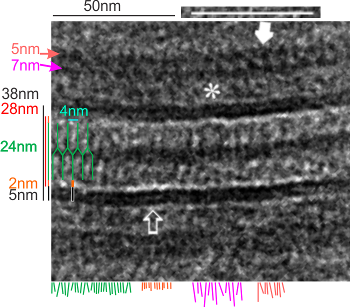
5nm approximate thickness of the cell membrane (one on each side of the desmosome shown here)
38nm from plasmallemma through to plasmalemma of second cell (THEIR MEASUREMENTS LIKELY TO THE CENTER OF THE TRILAMINAR MEMBRANE were about 33nm — pretty close)
28nm extracellular space (THEY DIDN”T MEASURE)
24nm is the @ height of the repeating units and N terminals of the desmoglein and desmocollin molecules (two molecules stacked=height (vertical dimension in this micrograph) (THEY DIDNT MEASURE). It is marked as the green stretchy-wire lines (likely a significant configuration for movement of cells that allows for some “give and take before break” which would not be surprising, but actually be awesome) and was easily drawn over many portions of many micrographs (this, and others of my own that i have posted before) .
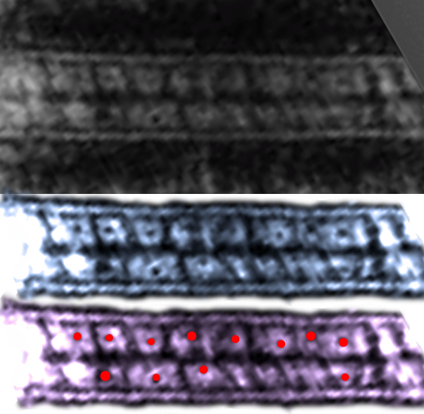
4nm spacing between the repeating but alternating units of the desmocollins and desmogleins (THEY THOUGHT THAT THE VERTICAL LINES WERE 5nm APART, I think more like 4nm apart and staggered).
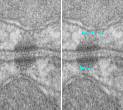
2nm orange lines, the lucent region just before the transmembrane segments of the desmocollins and desmogleins.
7nm pink arrow is a measured periodicity of one band of densities in the inner plaque, and the orange arrow represents measurements from adjacent periodicities.  To me they didn’t look the same… the latter perhaps being further spaced and still alternating. White asterisk is from the original micrograph in the Al-Almondi paper, outer white arrow points to the same group of proteins as my orante arrow labeled with 5nm distances. Their white outline arrow points to a lucent area that i did not measure which is just intracellular to the inner leaflet of the plasmalemma.
My measurement lines are shown below the micrograph. Green: (i should have made black since they were measures of plasmalemmal thicknesses).

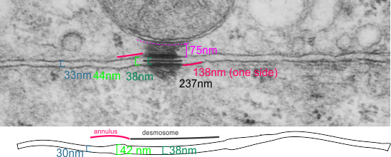
In liver, the intercellular space between hepatocytes is actually greater than the extracellular space of the desmosome filled with the cadherins….  which really is a very rigorously attached and spaced area.

One other difference in the way that Al Almoudi describes the “inner dense plaque” (most medial area of the intracellular desmosome structure) as having a single periodicity…. but when i counted distances between periodicities they actually were statistically different (p=0.03) with the most medial band of periodicities being further apart than those just adjacent and closer to the plasmalemma (but both in the inner dense plaque.

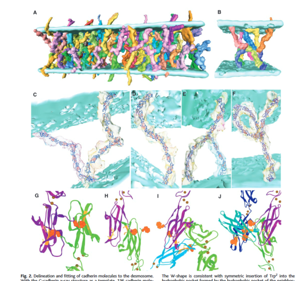
If i would going to hazard a guess on the shape of the molecules that make up the intercellular space of a desmosome i would have one which has a “blump” before the transmembrane domain, a blump at the N terminal (which is an obvious feature as the central periodic line in the desmosome).  The transmembrane part would be quite thin since there appears to be a lucent line just on the outside of the plasmalemma of the two adjacent cells, and before the linear densities are obvious. The current protein databases make an extracellular domain 3-D molecule for desmocollin and desmoglein have a good chance of being fit to the densities found in electron micrographs.