One transmission electron micrograph gave me a particularly interesting view of the intracisternal protein contents of a type II cell which has the large bodies filled with what I think is an organized layering of surfactant protein A. This tangential section was unique in that I could find many little objects which comprised a central density and six densities surrounding it…. making it pretty interestingly similar to the “bouquet” style protein 18-mer that is touted to be representative of surfactant protein A.
I have cut and pasted out ribosomes from the membrane of that RER profile for an approximation of the size (which is pretty clearly like other ribosomes at 20 nm in diameter) and kept the sizes relative to these circular hexagonal-with-central density areas in sync. You can see that they are just slightly larger than the ribosomes themselves, and that the estimated size fot he SP-A molecules (also given at about 20 nm) might be within tolerable limits.
The top rows of the image below are ribosomes and these circular proteins from a single tangentially cut profile of RER (cisternal body) from a guinea pig type II cell; the bottom set is from a not quite as nice tangential section of ribosomes and the circular proteins from a cisternal body of a ferret type II cell.
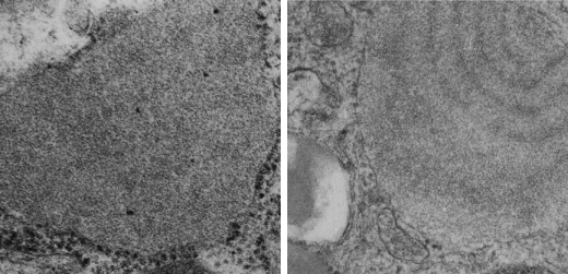
Below that image is a low res pair of images (guinea pig on the left ferret on the right) from which the ribosomes and SP-A? were selected.
I included one ribosome adjacent to a round SP-A ? molecule that has its own bar=20 nm.