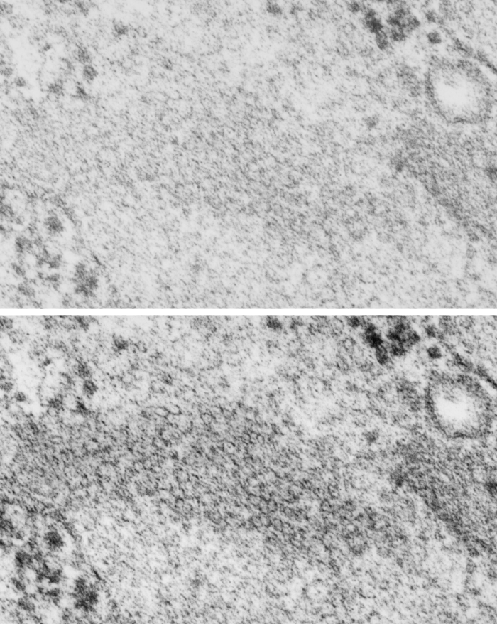
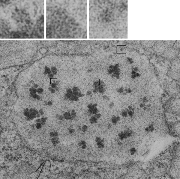
Dark band of an intracisternal body (dark bands in a perpendicular section of these intracisternal bodies span about 100 nm, but when spread out over a tangential cut, can have many more nm from the next dark layer in these inclusions. (I am calling them inclusions for want of a better word, but are rather sites of over-production and layered organization of what I think is SP-A. It provides an alternative view of the very orderly periodicity (3,5,7,9 – depending upon orientation and how thin the section is). These two images are identical except that I have increased the contrast, and burned (photoshop) the order which I see in these tangential portions (which likely represent the carbohydrate recognition domains of SP-A. I thought at first these hexagons were SP-A, then began to wonder if i could find similar structures “anywhere” in “any section” as an artifact of fixation, so went to several micrographs and scanned and examined them for randomly occurring small octagonal structures about 25 nm in diameter. There are many, but then I used the “light” banded area just as a control to the dark banding in an RER profile though to contain large amounts of organized SP-A as an internal control, and to help rule out the possibility that the fixation turns all proteins into patterned molecules (which it probably does to some degree) but which would fool one into thinking it was SP-A. So the bottom line is that I think the hexagonal structures (about 20-25 nm in diameter (see ribosomes nearby for comparisons in size at 20 nm).