Monthly Archives: June 2016

Nuclear pore micrograph and nuclear pore model

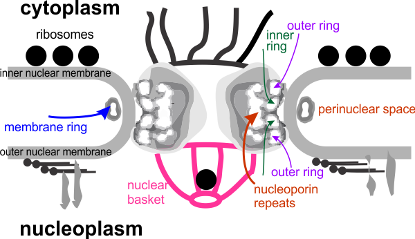
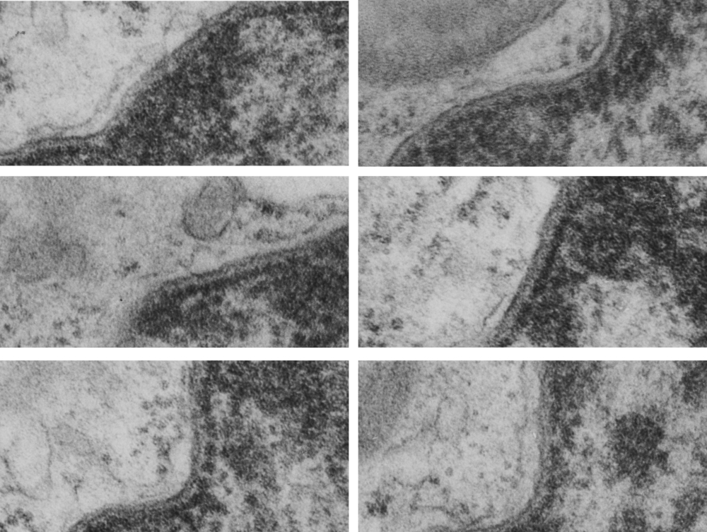
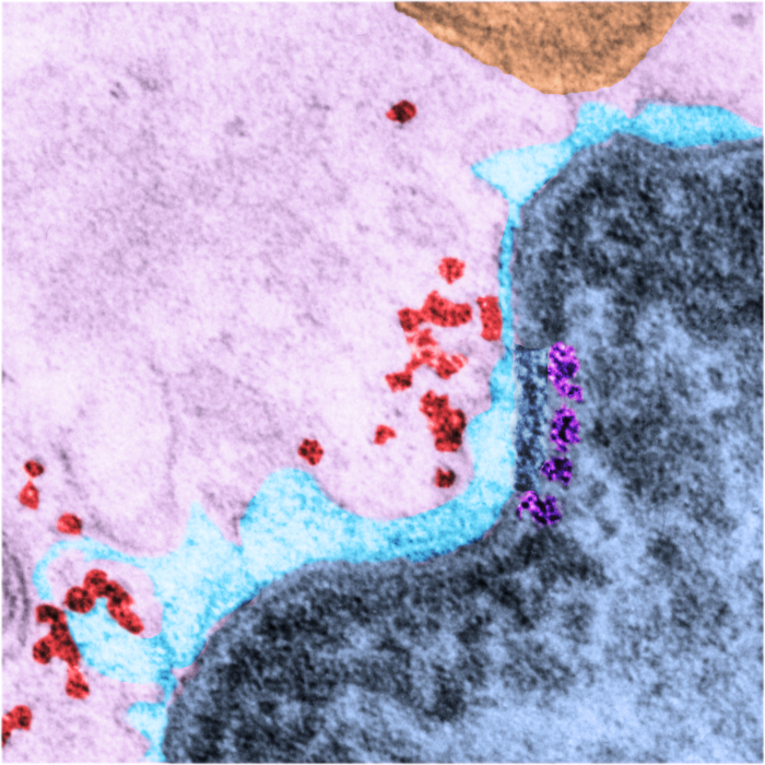
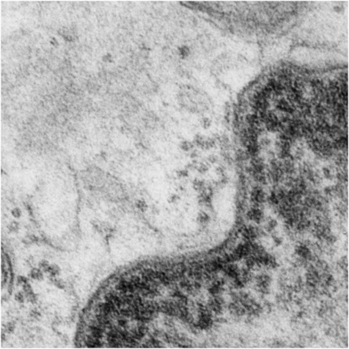
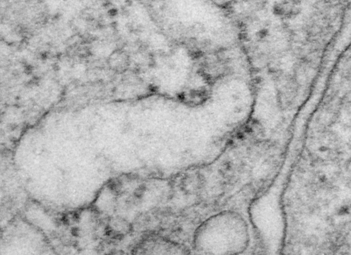
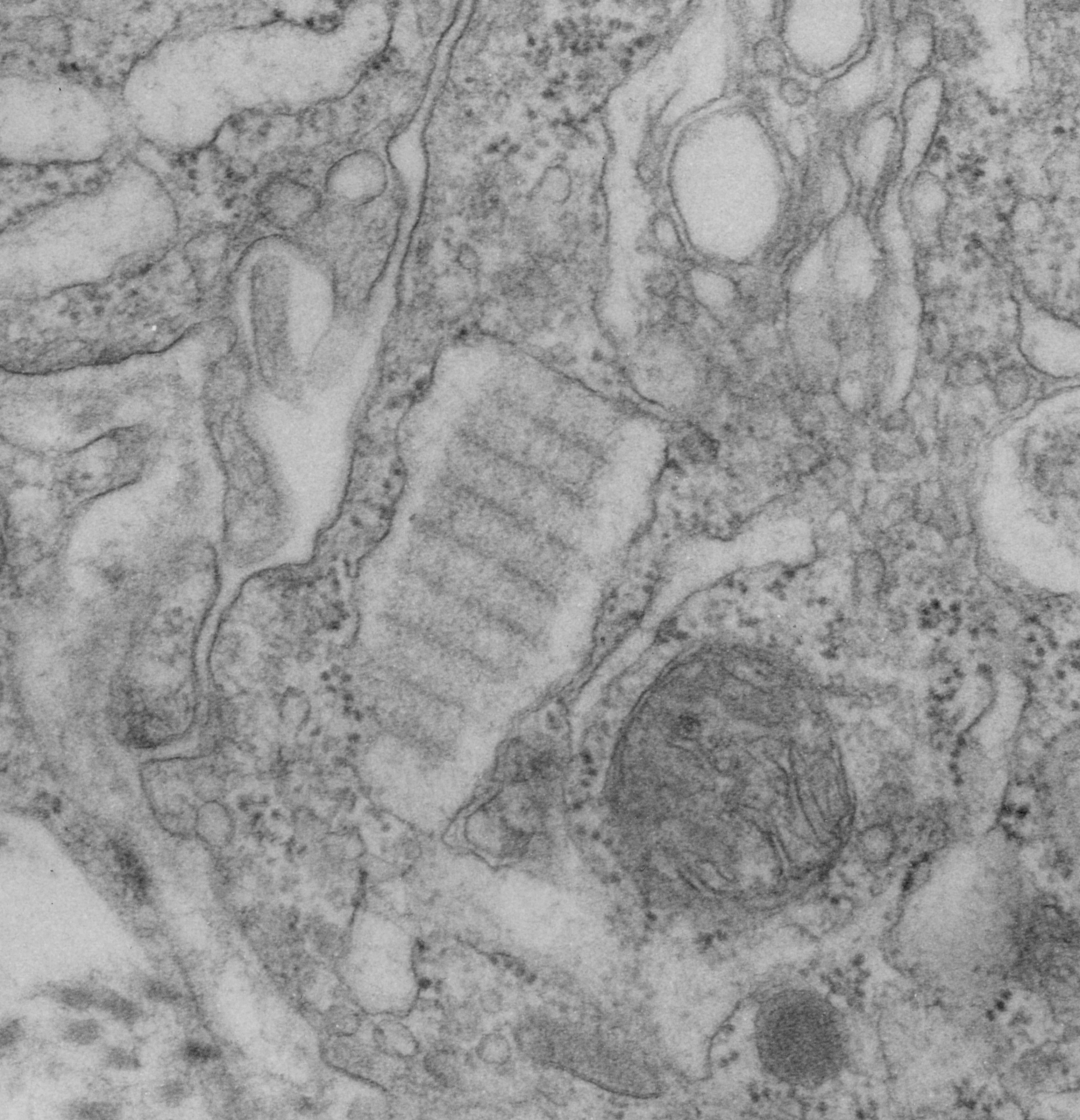
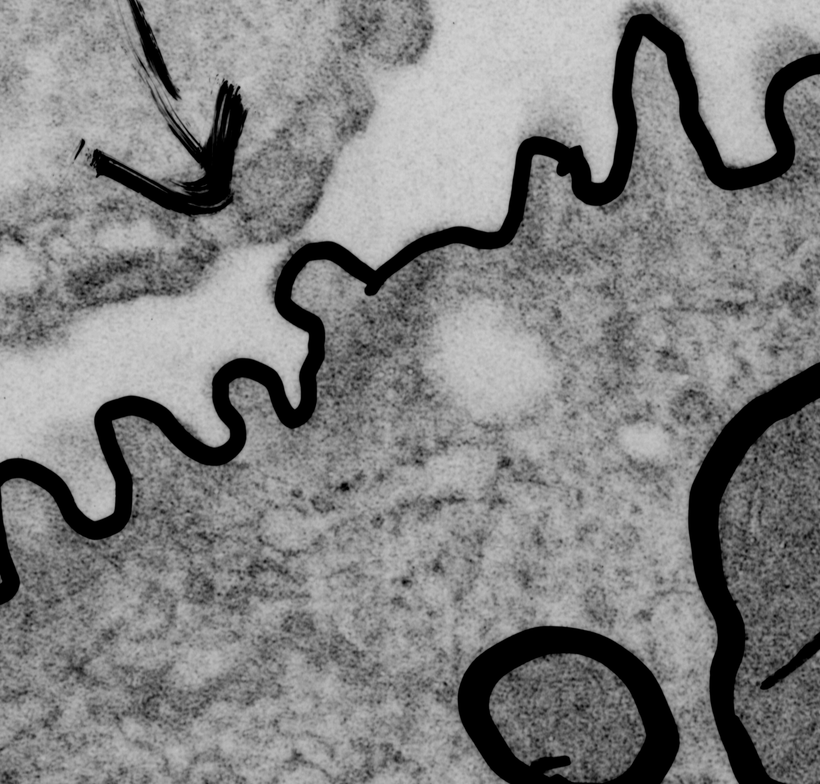
Yesterday’s post on a nuclear pore from a type II alveolar cell i pursued a little more, trying to establish a relative size for the current image and models of the nuclear pore complex, and a website posted this terrific image which i made transparent and placed into the transmission electron micrograph of  of a nuclear pore from my studies.  There are some issues using their image which is a top down view, and my image which is sectioned more from the bottom up, but it is an interesting match.  One thing i like about this particular model as opposed to many of the diagrams seen online is that it does NOT extend the nuclear basket too far into the nucleoplasm and have the inner and outer nuclear membranes too spaced apart in the area just adjacent to actual pore.  It appears in my micrographs as if the cytoplasmic filaments are greater length than any basket structure seen on the nucleoplasmic side.   So this more compressed model (fading in and out of my micrograph below) works best.

website where i found the image of the nuclear pore is here: http://phys.org/news/2015-06-high-performance-microscope-pores-cell-nucleus.html#nRlv

Verge of a dream: I think it might be bliss

keys_staffI think it might be bliss
as our eyes can’t quite adjust
as the dress rehearsal
lets out in the
phosphorescent light.
I think it is contentment
that is casual perfection
in strolling down
the streets of yards
that bloom without a
thought to end yet.
It’s just a guess or story
we tell to
make their lives
a romance though we
see them
only in a window.
Little thought is given
As the open
club door
lets blue notes carry
their way to freedom.

rlb 06-30-2016

Nuclear pore: canine type II alveolar cell

nuc-pore__dog_type_II_cell_nucleusThis cropped portion of an electron micrograph of a nuclear pore was taken from the nucleus of a canine type Ii alveolar cell. It shows the filaments on the cytoplasmic side, and a bunch of ribosomes positioned just outside the nuclear pore, and two very distinct structures, about ribosome size that are either coming or going through the nuclear pore.  It made me laugh, as it almost seemed as if they were going or coming in a “pin ball” fashion, being shot through the pore directly in the center. Do you see both of them? one is almost dead center of the micrograph, abd just elow the nuclear pore, and one is almost directly below that.

This electron microscopic image prompted me to try to figure out what is known about the nuclear pore complex in general, and every diagram I found in the literature was just a little different, even that found on wikipedia had some elements that I could not assemble over top the micrograph (which is basically what i wanted to do), so the diagram which accompanies this fine structural view (ultrastructure) is way off… I would love to find the time to put together a better diagram.

nuc-pore_diag


Organization of chromatin along the inner nuclear membrane

Protein complexes that are involved in the regulation of transcription might be located near positions on the inner nuclear membrane. Certainly, at the very least, the architectural proteins (lamins) are involved in this, and the lamins which are present on the inner nuclear membrane may be as well.  Yesterday’s post revealed something I had not seen before which made me curious about the different (I saw at least three easily) “stringing” patterns of chromatin on the inner membrane, one very loose, one quite tightly together, and the third somewhere in the middle.  I searched about 100 other micrographs of type II cells from three species (ferret, dog, guinea pig) and found many examples of the tight configuration.  Accompanying the tight configuration is an apparent rigidity in that particular stretch of the inner nuclear membrane.  Searches online did not reveal much in the way of literature to explain it.  It seems reasonable to assume that the tightly spaced areas of DNA and other proteins is not going to be active. There were occasions when the tightly packed chromatin was close to a nuclear pore, but not always.  It also seemed as if there was a slightly greater electron density to the lamin area just beneath the tightly packed row than under the medium or loosely packed chromatin.

Red arrows point to loosely packed chromatin right at the inner nuclear membrane adjacent to the lamins, white arrows point to the tightly ordered chromatin, also right at the inner nuclear membrane adjacent to the lamins. Nuclear pores, pink, nucleus, blue, nucleolus purple,  lamellar bodies, yellow, cytoplasm, green.  There is also in this photograph a mitochondrion (the green color of the cytoplasm, in the lower left corner which is very close to the filaments of the nuclear pore….. something which is quite common and also something which I have not seen explained.

9933_24669_dog_type_II_cell

Three obvious states of inner nuclear membrane organization

Just looking at a single nucleus, single inner nuclear membrane (INM), single cell, it becomes quite clear that the organization of the INM varies and that three very prominent differences in that organization are apparent.  A normal configuration (I am only calling it that because it is “in between”, and a very dense organization, and a loose and spacey organization (see previous post on june 24 2016).  The image below is something of a quick and easy demonstration of that point. Electron micrograph of the inner nuclear membrane was cut in six different places from an image of a dog type II alveolar pneumocyte, a control animal from experiments decades ago, and It was easy to see three types of organization.  tight, middle and loose.  It will be interesting to determine which processes go along with which anatomical arrangements (though the outcome is not difficult to predict).

Top=two segments of the inner nuclear membrane which is  “neither tight nor loose”, and orderly; middle row=two examples of “tight” organization and bottom row, two examples of “loose” organization.  Inner nuclear membranes all coursing upper right to lower left, condensed chromatin is on the right hand side of the nuclear membrane, none of the profiles contains a nuclear pore, the slightly less dense layer of lamin is present in all images, some areas of euchromatin are seen, upper right image has a portion of a lamellar body, two other images each have a portion of a mitochondrion, scattered ribosomes are on the left hand side of each image, and the perinuclear space is not particularly dilated in any of the images.

loose_tight

Transmission electron micrograph: nucleus

The nuclear membrane is interesting, to say the least, the perinuclear space the “literal outside” and the two bounding membranes fluid and changing positions in an active and rarely appreciated way. I can remember in the late 1960s working at a place called “Pasadena Foundation for Medical Research” which no longer exists, watching some of the first and best cellular time lapse photography, the nuclei spinning and jerking around inside the cell, mitochondria dancing close, touching the outer nuclear membrane (in what I called nuclear pore-mitochondrial interactions in some paper long ago.

There are dozens of important proteins as part of the inner nuclear membrane, organizing and facilitating every aspect of transcription, translation, expression, silencing, condensing, organizing, modifying nuclear DNA and RNA and all components therein the likes of which I know practically nothing.  I do know the organization of the inner nuclear membrane visually…. and this little segment of proteins – slightly larger it seems, than a ribosome (or about 40nm) is special!  It is pseudocolored magenta, and it has ordered and deliberate spacing not seen normally (check out the rest of the inner nuclear membrane adjacent to this stretch).  The nucleus itself is colored blue, and the perinuclear space, light blue, ribosomes in the cytoplasm are pseudocolored bright red-orange, the mitochondrion is orange and the cytoplasm is pale pink.  Top image is pseudocolored, but the bottom image is black and white, unretouched from the original micrograph (canine, 5 yr old, untreated, alveolar type II cell, lung)

9933_24669_dog_INM-color9933_24669_dog_INM-bw WIKIPEDIA says “There are about 60 INM (inner nuclear membrane) proteins, most of which are poorly characterized with respect to structure and function.[2] Among the few well-characterized INM proteins are lamin B receptor (LBR), the lamina associated polypeptides (LAP1, LAP2), emerin and MAN1″  italics are mine, sentence shortened as well.

HERE is a link to a nice diagram and article of the inner nuclear membrane in terms of the currently known proteins. http://www.nature.com/nrm/journal/v8/n5/box/nrm2165_BX1.html

Rough endoplasmic reticulum, ribosomes, and protein synthesis

Pseudocolored electron micrograph depicting what I think is 4 separate actual protein transcription events within an RER profile — see as four ribosomes on the RER membrane and the resulting 4 strands of protein within the lumen of the RER. The RER cisternal profile is pseudocolored cyan, the ribosomes (4 of them in particular) are blue, the proteins being synthesized, 4 of them, from adjacent ribosomes are in red, and the extracellular space beside the type II alveolar cell (from a ferret) is brownish, and the type II cell cytoplasm itself is a light violet color. This was an accidental find while perusing hundreds and hundreds of type II cell electron micrographs from a half dozen species of mammal.

This particular micrograph was not taken FOR this purpose, but it illustrates a point in time frozen by fixative and plastic three decades ago.

9777_34065_RER_ribos_prot_color

Text book illustration electron micrograph: RER, ribosomes, protein production

This electron micrograph just made me chuckle because for whatever reason the fixation of this particular profile of RER just had four cute little ribosomes lined up and stringing out from them into the lumen of this profile of RER were four strands of protein, even to the point of having a little kind of fat area at the leading end….

Here is a contrast enhanced (I did use the burn tool in photoshop on the ribosomes and the protein) portion of that ferret type II alveolar cell, and below that is the area from which I cropped the profile of RER.  Magnification and enlargement can be approximated from the ribosomes… which are @ 25 nm in diameter.  Below that is the original micrograph, not retouched.

9777_31065_ferret_ribosomes_protein_inset9777_31065_ferret_ribosomes_protein9777_31065_ferret_ribosomes_protein_untouched

Ferret type II alveolar cell RER showing parallel bands in organization of intra-cisternal protein

How awesome is this particular view of an intracisternal protein in a ferret type II alveolar cell. The bands are slightly tangential and so only the most electron dense banding is seen, however, the interesting thing here is that on the periphery there is not really any banding, but it looks as if the banding occurs centrally in the granule (aka within the profile of the RER).  Also, in the upper left of this electron micrograph (in an adjacent type II cell) examine closely the profile of RER  which actually can be perceived as ribosomes on the membrane surface and the trails of protein hanging off in the RER lumen.  Really a classic text-book presentation in “real life” so to speak, though we all know that TEM captures only a “nanosecond in time”, and all proteins are “fixed” and so distorted. Nevertheless, this may be one of those opportune views.

The protein (which I think is SP-A) is central in the micrograph, the banding pattern is at about a 35 degree angle.  Ribosomes are present on right and left borders for the most part, smooth ER is in the upper right corner and there is a tiny portion of a lamellar body off on the right side.  A portion of intercellular space with a couple of plasmalemmal folds crosses from top center to bottom left. There are a few other profiles of RER without SP-A? banding as well.

9777_31065_ferret_crop

Cat: periperhal lung type II cell

Lung tissue harvested from a sham treated control from another investigators experiment (thereby saving time – money – animals) here is a piece of a type II cell viewed with the electron microscope. It has my markings (for morphometry, back in 1982) and a marker arrow pointing to an area of RER which was the closest thing i could find to an organized protein in the RER (as a continuation of the search for species which may have SP-A organization similar to that found in guinea pig and dog and ferret.  In cat, though the total number of electron micrographs I have saved is not that great (a dozen or so) therefore the absence of such a layered intracisternal protein is not to be assumed).  The ribosomes studding the RER membrane provide an estimate of size (each being approximately 25 nm in diameter – a general number for all species and conditions…. (generalities are not always good for much, but an approximate size is OK here).

7296_24554_cat_3_lung