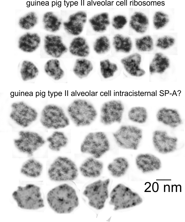
Still working on whether this is SP-A (surfactant protein A) in the layered protein paracrystalline type banding seen in the RER of at least three species of mammal. The most prominent of these banded protein products in the RER of alveolar type II cells seems to have occurred in guinea pigs, though they were first observed in ferrets. These six images are high mag images where just the RER with the layered protein are highlighted (with the black arrow). Since they all came from different images and magnification is just to messy to calculate I have substituted the 20 nm ribosome as a “scale” so the red dots are a “relative” measurement of 20 nm. This puts all the intracisternal bodies in perspective.
The dog is different than the other two species in which the intracisternal body in RER of type II alveolar cells is seen as I have only found them with a single banding period…. whereas in guinea pigs the number of periods in a single RER profile with this protein can be very large and difficult to count because of the perpendicular and arched and oft changing directions of the layering. However, the mean number of bands in guinea pigs, 787 periods, n=189 RER profiles, produced a mean of 4.1 (SD=2.9) +- 0.21 (SEM) periods per profile while in ferret of the 625 periods counted, n=123 profiles of RER, the mean number of periods per profile of RER was 2.66 (SD=4.9) +- 0.39. Mongrel dog had very few profiles, and number of profiles was = to the number of periods (n=7). Six of those are shown in the figure below: they were about 100 nm thick and maybe twice that long.