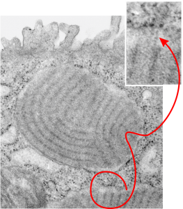
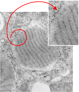
Electron lucent areas just beneath the RER limiting membrane alveolar type II cell, these have ribosomes and layered protein, just like the previous post. I suspect it has to do with the proximity of ribosomes to the protein beneath, and the lucent areas just seem not to have close-by ribosomes. The red circle highlights which part of the intracisternal body is being noted, and the arrow head points to the insert and enlargement of that arrow. As before, the original large image is unretouched while the inset has been “burned” in the area that I want you to see, which includes the more electron dense area and adjacent more lucent areas, and the ribosomes. This is an alveolar type Ii cell from a ferret, magnification is 19,500, enlargement before scanning is 4 x. Final magnification can be more or less approximated by the size of the ribosomes (reportedly 20-30 nm) which makes the dark bands within the intracisternal body separated by a distance of about 100 nm.
Daily Archives: July 27, 2016
Electron lucent areas just beneath the RER limiting membrane
 Electron lucent areas just beneath the RER limiting membrane seem to be a feature of very large and complex intracisternal bodies (ICB) (or RER SP granules if you prefer to give them a name that is what I think they may be (that is surfactant protein granules, backlogged in the RER for some reason)) This particular ICB has all the earmarks of the same layered striations seen in guinea pig and dog (this is from ferret, and is way more abundant that typically found and more curvey as well, 6911, 23303 19,500 original mag, 4 times enlargement plus whatever playing i did in Photoshop to crop and enhance contrast).
Noted here is the pale area beneath the cisternal membrane (RER membrane) which waxes and wanes around the central layered protein area which is considerably more dense than the pale areas. And then in occasional areas where ribosomes are present on the outer edge of the membrane, there can be some streaky connections (see inset) and a little bit of increased electron density in that electron lucency. In the inset the ribosomes, 3 of them, and streaks, 3 of them, have been “burned” using photoshop… the larger and original image was not “burned” in that area and the densities can still be easily seen (red lasso around the area that is enlarged for the inset which lies at the end of the arrow tips).
