Monthly Archives: August 2016

Nucleated red cell collage

Anatomy is so awesome. The order is fantastic. Our inability at this juncture to understand the massive amount of detail involved in just the function of one single nucleated red cell is overwhelming. Here is such a red cell, electron micrograph originally, which I have pseudo colored, cut pasted,  filled and framed.  Just look for the mitochondria and vesicles, maybe a little RER in the middle and lower left, and hunt up the nuclear pores and look for the areas condensed and euchromatin, then smile.

nucleated_red_cell_flower_collage

Kroger Vegetables and Noodles: or should it be Noodles mostly.

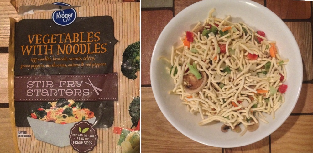
One of the things in life that I really find unpleasant is when corporations, people, universities, children and spouses just feel the need to “juice” things up over and above what the “reality” is.  The packages of mixed vegetables and noodles made (distributed) by Kroger galls me every time, but tonight took the cake.  If there are MORE noodles than vegetables in the package (by weight, by volume, by whatever criteria you need to use) then don’t name the frikkin product…VEGETABLES and NOODLES…name it NOODLES and VEGETABLES.

I almost never open a package of this stuff that I don’t feel some disgust for false advertising.  Why do consumers put up with it. Why don’t corporations have a “soul
or “morality”, why is capitalism gone “big” as in Kroger, K-Mart, Walmart, Verizon, Duke energy, Novartis, Pfizer, GlaxoSmithKlein, and so on ad nauseum …. take license to cheat  customers.

I don’t need to belabor this, we can change the world, one blog, one phone call, one boycott, one choice at a time.

These pictures are “AS IS”,  just cropped to a same height.– I had just emptied the whole package into the white bowl seen, and thought to myself…. tonight is the night I post this lie for the jillions of people who know what I am trying to say.

kroger_vegetables_and_noodles

Rabbit alveolar type II cell: is this surfactant protein A or not?

Going through additional species of animals looking for possible corollaries for the intracisternal bodies found in the RER of guinea pig, ferret and dogs, I examined just a few transmission electron micrographs of rabbit. One animal from a study with Leland Clark Jr, very likely involving artificial blood, and four other rabbits in studies with David Warshawsky, with carcinogens (likely dibenzcarbazol, and also iron oxide).  In the former (see the unretouched, but contrast enhanced micrograph below) I did find three stacked RER cisternae which had the look of some of the cisternal bodies found in three species but I was not able to detect any of the layering–aka—periodicity within these cisternae.  Well actually i was tempted to see it, it seemed to me that very faintly there was some tendency to see banding.  You can judge for yourself. Top micrograph.

In the bottom micrograph at the thinnest part of the cisterna (inset marked by a black rectangle and enlarged to upper right corner) at a near perpendicular cut, it seemed to me that the cisterna was more than 100 nm in width.  (A ribosome adjacent to the cisterna was used as a measuring stick, the latter presumed to be 25 – 30 nm in diameter;  red dots). What might be construed as linear patterning, or parallel periodicity, appears as red lines.

11320_37150_rabbit11320_37150_rabbit1

Amazing! and not to bright.

I have worked in environmental health for almost 40 years (well maybe a little more if you count postdoctoral training). And most if it has been in the same department. This says much about my resistance to change, but it says nothing about my desire to “care”
and “spare” when very old buildings are thoughtlessly removed, renovated, revived etc.  And I am not against progress, but I am also not for change for the sake of adding new names to new facades that are no better than the ones erased.

I was always amazed and provoked and saddened working in environmental health because it was a “tag” for most workers here, not a passion.  We still had boxed lunches in ugly wasteful plastic containers, recyclable cans thrown into the regular trash, no plan for reminders when staff took their recyclable papers and cardboard out to the regular dumpster. No thought for the escaped coolants, or energy conservation (making some parts of the buildings too cold to work in without a jacket, others so hot and dusty that you suffocated using the stairwells).  Even cleanliness was spotty in the buildings (not good when there is lead paint on every wooden window, literally everywhere, toxic dust, ….  some asbestos, traces of other things left over from experiments…..and where I workeugg_emogid for so many years was often left on the “outside” .  Ha ha… My coworker (unnamed for their protection) and i used sharpie to draw a circle around a large dead insect which sat on the back stairwell for “literally, not figuratively” years.  OK, so i am not being a grump…  but now the Old College of Medicine…. not the one built in 1972, but the one which housed pretty much all the medical departments for basic science for the first two years of medical school in the “before time”… which has been renovated a couple of times. I remember one such 3,000,000 project which raised eyebrows, now obliterated by another iteration…. is being power washed.  This in and of itself is not a bad thing, but two men spraying down windows most certainly covered in chipped old lead paint are doing so with no respirators, and putting all that filth into the neighboring ground… hmm  right next to a hospital… tracked into many other buildings, and even the home of students attending the college of medicine, and the department.  Something doesn’t work here.  I am more than happy to take your comments if you can prove I have spoken out of turn…..  your questions, though, will have to find a way to me besides the “comments” section on this blog, since that is just a place for selfserving spambots.

Biggest alveolar type II cell RER granule so far

Guinea pig (now the most posted alveolar cell-RER granules (intracisternal bodies) in history (LOL) guinea pig # 301.  This is a very very large protein accumulation, and somewhat different in morphology than the typical intracisternal body (seen adjacent, below) and I don’t know yet why the difference in structure.  This guinea pig, that is, the guinea pig 301 which continually showed the greatest number of RER granules of any other animal in this archival set of micrographs, and interestingly it possesses some distinct prismatic (rhomboidal) regular bodies that resemble virus particles (more data needed).  It would be awesomely consistent with what is known about surfactant protein A and innate immunity to have this guinea pig demonstrate first hand what kind of an increase in surfactant proteins (either A or D) to respond to a viral infection.  We will see.

This granule rivals in size the nucleus in the adjacent alveolar type II cell.

9817_17084_gpig_301_typeIIcell

Osmium tetroxide fixed alveolar type II cell, dog, intracisternal body

Going through archival electron micrographs of alveolar type II cells has been an interesting study. The purpose of the review is to gain insight into what is the explanation for some very interesting, highly organized granules (protein accumulations) that are found in some alveolar type II cells.  Of about 8 species examined so far (rat, mouse, owl monkey, rhesus monkey, dog, ferret, guinea pig, hamster  – even one human case) these bodies have been found in only three species. Dog is one, though in dog they are quite rare, and very small and not nearly as developed as found in ferret and guinea pig.

Most of the archival electron micrographs are tissues fixed in a double aldehyde fluid, in various buffers and at reasonably constant concentrations of glutaraldehyde, around 2.5%.  This tissue however was fixed in osmium tetroxide… thus verifying pretty much that the cisternal body granules are NOT just an artifact of aldehyde fixation.  So this image of a dog alveolar type II cell is not that great a micrograph, but it does show a small, typical style dog intracisternal body granule, with a similar periodicity and banding as found in aldehyde fixed material.

Upper image shows the mostly unretouched (i changed contrast, and took out a few scratches but did not change any data) and the bottom is a highly contrast enhanced crop of the upper image, and i have used the burn tool in photoshop to highlight the periodicity of the middle band (as also seen in many other micrographs posted in this blog).  The ribosome standard I called 27 nm in diameter, which makes the dotted periodicity of the central band of the cisternal body about 17 nm in diameter (also shown as a red dot) and the space between those 17 nm dark protein dots is a distance (made from 12 adjacent measurements) a mean of @ 32 nm.  Band across the body is close to the 100 nm that has been found in other instances in ferret and guinea pig and is presumed to be a collection of some multimer of surfactant protein A.

dog_osmium_fixed_icb


Alveolar type II cell morphometry: old school

This archived electron micrograph is a sample of the old school style of morphometry. Here is a ferret alveolar type II cell which, when printed, was overlain with a scored plexiglass grid during photographic exposure, to make it easy to count intersection-crossings to determine the volume density of organelles, such as mitochondria, lamellar bodies, RER profiles, and also to count intersections of linea profiles for organelles like RER and plasma membrane and nuclear membrane.  You can see in this very old sample that I circled the nuclear pores.  Cross marks at intersections counted are still visible (ink lines).  Though this is seemingly primitive, to me it allowed much more precision that using image processing, since the eye can adjust to differences in light and darkness in the prints, and pick up debris and other extraneous junk and scratches that might just be counted as “objects” in computer vision programs.  Besides being more flexible, probably more accurate (my opinion of course) it is also quick, and does provide a “hard copy” to go back and verify and spot check one’s data (which counting on a computer doesn’t always do. Even more importantly, not all information that is desired is known at the time of sampling, and therefore having a resource to check back with is invaluable.

4532_18332_ferret_typeII_morphom

Contributions to mutations from male and female (stained glass pattern !)

I read a post from the Center for Environmental Genetics (I run the blog for this man (a good friend) who searches the literature for great articles on gene environment interactions  http://genewhisperer.com ) and it mentioned the relative contributions that are believed to arise in the new generation as determined by the different contributions from the parents.   It made me really realize that things are divided…. ha ha that sounds like a simpleton, but it is so clearly a binary universe, oughts andmale female variation_LRG ones, female and male each providing input on the de novo mutations in this case the number of divisions for a new sperm cell ( now as many as 640 divisions by male age 40) provides so many chances for error, and the femal has eggs at birth, …. no divisions in the femail egg so the de novo mutations here are quite small.  HOWEVER, that said, other factors can derail the system….epigenetics for sure, and maintenance of the DNA, repair errors etc.   It is soooo complicated, that even in 100s of years, it will not yet be known. This stained glass pattern can be found HERE.

So this stained glass pattern is the yin yang, up down, male femail, september october, combined forces pattern with just some really great lines, and curves, the female and male signs from astrology, alchemy and modern science combined.


Nuclear portion of an alveolar type II cell enlarged to show nucleoplasm organization

6760_17082_gpig_nuc_pattern

Looking at an electron micrograph of a guinea pig alveolar type Ii cell, (my guinea pig number is 301 – block 17082) archival material from the 1980s, I found this really interesting pattern in the nucleoplasm just above a nuclear pore.  On either side of the outer nuclear membrane spanning the nuclear pore was the granule protein stuff that I have been working on, trying to place it possibly in the surfactant protein family, but being overproduced to the extent that it becomes its 18-mer, in mirror and/or vertically flipped position, times 2… producting the 100 nm banding that I see.  But for this nucleus, just to the right of the nuclear pore marked with a two headed arrow pointing to the lime green intracisternal protein within the RER, there is a pattern that just “popped out” to me which is quite large (relative to the size of a ribosome…. and NOT to be confused with the hexagons in a tangential view of a granule but in the NUCLEUS proper) and sort of hexagonal in shape (which is what one expects when pressing together spherical bodies which I pseudocolored a salmon peach.  Red circles surround structures that look like perichromatin granules (I am sure I could have found more).

Intracisternal granule from alveolar type II cell of the guinea pig

Nucleus, other organelles, nuclear pore, intracisternal protein accumulation, perichromatin granules, and part of the basement membrane are labeled in this electron micrograph of a type II cell from the lung of a guinea pig. Nucleus is on the right and a very thin band of cytoplasm is present, then there is the extracellular space (with basement membranes and parts of other cell processes).  Basement membrane – purple arrow and label; red arrow and label – nuclear pore; black arrow and label – intracisternal protein which might be surfactant protein A (overproduced). Perichromatin granules (adjacent to heterochromatin) are shown by blue arrows and label.

6760_17082_301_gpig