Daily Archives: September 2, 2016

Microscopy Today: Cover for January 2008, apoptoses as snowflakes

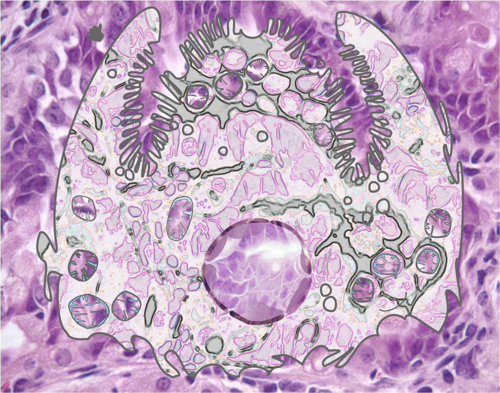
This was a cover that I had hoped would be published with an article that I helped to co-author that described a pro-survival gene, or otherwise functioning as an anti-apoptotic gene.  Playing with the images of apoptotic cells in culture I did manage to submit the image to Microscopy Today, and they published it in 2008.

I thnk what amazes me is that I really made a whole lot more cover submissions than were successfully published (LOL). I find a similar statistic in publishing data, where really only a small part of what is studied, learned, documented, and put out there in the world of science, makes it into print.  Good things have to get strained through the teeth of overworked and sometimes not that interested peer-reviewers.  Who, I wonder, thinks so highly of themselves that they have the right, and/or mission, to exclude the works of others.  Notwithstanding the well know fraud in science, the great majority of individuals working to find “truth” in all its glory, are honest and well-intentioned.

snowflakes_apoptosis_Microscopy_Today_jan_2008_

Cover submission to JBC which was a finalist but didn’t make the cut

How awesome is this composite of gastric epithelium (light micrographs) put together in a collage of images with the forestomach images in the upper right outside, and the glandular stomach on the outside lower left and bottom and all the glandular stomach epithelium at lower magnification with light microscopy surrounding two higher magnification (100x at least) images within the center.  Too bad this didn’t make it on the cover of JBC.  Here is a mockup of what could have been.  LOL.   Still a great image.  I have to consider these cover rejections as just part of the competition in scientific illustration.

A7_A9_cover_draft_for_JBC_3

Parietal cell (stomach, mouse) with some organelles diagrammed

More images of gastric epithelium in the mouse, onto which is layered some vector graphics of mitochondria, vesicles, nucleus (pale condensed chromatin areas within, nuclear membrane (inner and outer membrane and perinuclear space — but no nuclear pores are identified here specifically but the places where outer and inner nuclear membrane come together nuclear pores would be found.  This is color and information together, for the 21 century and the digital age.

The microvillar (highly microvillar) apical membrane is shown, the lateral membrane and basal membrane are not distinguished from each other except as the bottom of the diagram is basal.

parietal_cell_pseudo-colored_with_diagram

Cardiac muscle cell and ion pumps diagram

This illustration, a background of cardiac muscle cell (mitochondria blue; myofilaments, myofibrils, purple) with an overlay of a rectangle representing a cell (no plasmalemma distinctions on this box) showing some of the ion pumps that are responsible for homeostasis and function in cardiac muscle.  This was a cover submission to Journal of Biological Chemistry, years ago, and unfortunately I had two cover submissions for the same month…. this one lost to my other one (ha ha). That was a banner month for me. Two submissions to the same journal but I just wish that either one or the other of the first authors on those manuscripts would have opted to delay publication one month, as I thought this was a pretty good graphic.

cardiac_muscle_ion_pump_cover_submission