Daily Archives: November 28, 2016

Hamster: alveolar type II cell

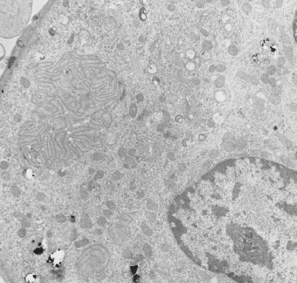
Another example of a species in which I have hunted from layered granules in the alveolar type II cells is hamster (though only a few animals have been studies). There are, as in cat and rabbit) frequent examples of RER which has a slightly dilated appearance, and also has obvious protein density within the lumen of the profiles, but lacks all but the very faintest appearance of layering, and also does not abide by the criterion for layered granules in that ribosomes stud all aspects of these RER profiles, and the rigid looking, ribosome free, part of the granules seen in ferret, guinea pig and dog, thus one has to assume either that surfactant protein A is sufficiently unique in the three species above where granules can organized into a regular substructure, or that something else altogether is responsible.  hamster, male, animal# 3, neg 7278, block 24733,

hamster_lung_type_II_cell

Cat lung: alveolar type II cell

Cat lung: alveolar type II cell, with arrows to three profiles of endoplasmic reticulum which have a structure not typically found in alveolar type II cells of common laboratory species (mouse, rat). These are rounded and it is not actually that hard to see some striations in them, particularly the one on the most left.  In a series of micrographs from untreated cats (taken incidentally from other studies) no sturctures with highly visible layering in the RER were seen, which doesn’t mean these profiles are not surfactant proteins, it just means that they are not organized as seen in ferret guinea pig and dog alveolar type Ii cells in other studies. negative 7268, 24564, cat #3, untreated (gender?, age?)

cat_lung_type_II_cell

Rabbit lung: post intratracheal instillation of iron oxide and dibenzo cabazole

Rabbit lung: post intratracheal instillation of iron oxide and dibenzo cabazole, electron micrograph. This is not a great picture, but i post it just so one can appreciate that there an area of odd membrane swirling in the middle left, and a small area just like it in lower left. The cell has a lobular nucleus and there are iron oxide (probably with DBA alongside) within this cell. I don’t think it is an alveolar parenchymal cell, but likely an inflammatory cell. A type II cells from the same microgrpah has lamellar bodies, which this cell does not, which further indicates it as a migratory cell. Animals given DBC do get tumors. neg 4447, block 17793, Rabbit #32-3.  Sorry the micrograph has stain ppt on it, but it is worth looking at just for the layered structure.

rabbit_lung_iron_oxide_dibenzo_carbazole