Monthly Archives: December 2016

Tangential cuts through alveolar type II cell granules show dense layer substructure

Tangential cuts through alveolar type II cell granules show dense outer layers of this regularly arranged molecular substructure, has a periodicity to it, albeit densities of a different size than at other sites in the , just like the less dense middle band of these granules.  Images below also show a slight tendency for vertical banding, within the horizontal layers of the surfactant protein (A ? ) granule. The hierarchy for all the level of substructural organization are here:

  1. 100 nm period (height) (no restriction on length of granule seen) which has 1,3,5 or 7 layers between the outer dense bands
  2. Outer dense layers on perpendicular cut look continuous, on tangential cut however, they show a periodicity with large round densities about 2.5 per 100 nm length.
  3.  Moderately dense central band shows a periodicity of 5-7 densities per 100 nm
  4. vertical stripes, a kind of banding, perpendicular to the long orientation of mos that coinciding with the periodicity of the central dense  band.

Negative 7088_23356)ferret_#17more_vertical_banding-1more_vertical_banding-2more_vertical_banding-3

Vertical and middle layer organization in alveolar type II cell surfactant protein granules

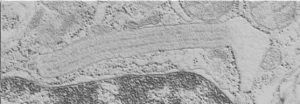
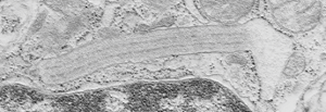
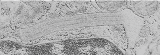
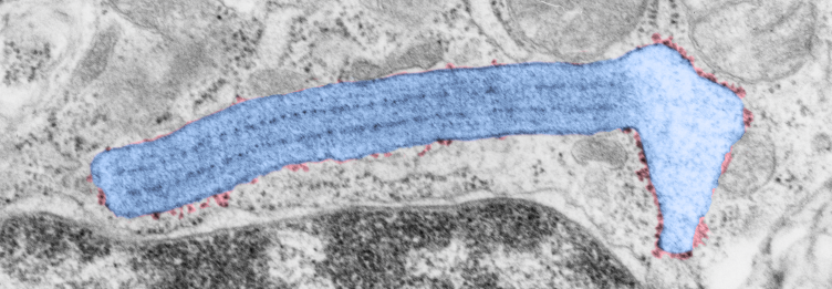
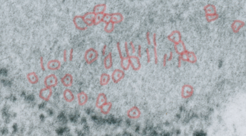
Vertical and middle band banding in alveolar type II cell surfactant protein granules is shown here (hopefully this is SP-A). Using the same image as posted as the quintessential surfactant protein granule in alveolar type II cells, the image has been copied, inverted, and offset just a few pixels, to cast a shadow and give dimension to the “edges” in the image, a kind of embossing technique.

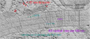
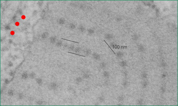
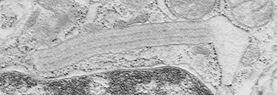
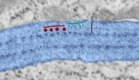
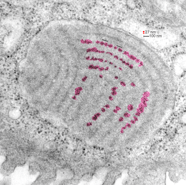
Embossing vertically highlights the bumpiness of ribosomes and other protein “lumps” within the granule, while embossing by moving the inverted image below, causes the ribosomes to appear to be concave.  Both accentuate different features (see below). The embossing upward as well as moving the inverted image a few pixels to the left highlight the banding seen within the granule which is secondary to the electron dense bands that run parallel to the long dimension of the granule… the vertical lines are not present in the surrounding cytoplasm, thereby indicating that the vertically lines within the granule are not section artifact or fixation artifact but actually a part of the specific alignment of the granule proteins.  The third image is a flattened image, cropped, increased in brightness and contrast, then vertical lines were superimposed where I saw definition.  Ribosomes, as an internal control for magnification are about the size of the red marker (27 nm estimated diameter), black line of 300 nm = the height of the three banding periods comprising this particular granule, 100 nm bars mark out the middle dense layer (with the blue dots where periodicity shows up) and also shows that about 4-5 protein densities occur in the middle layer of each 100 nm period.

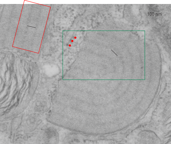
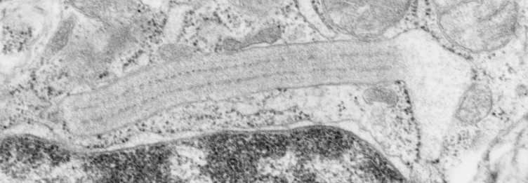
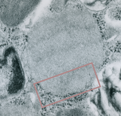
Original ferret alveolar type II cell RER granule electron micrograph, untouched, from which these processed images were derived can be found here.

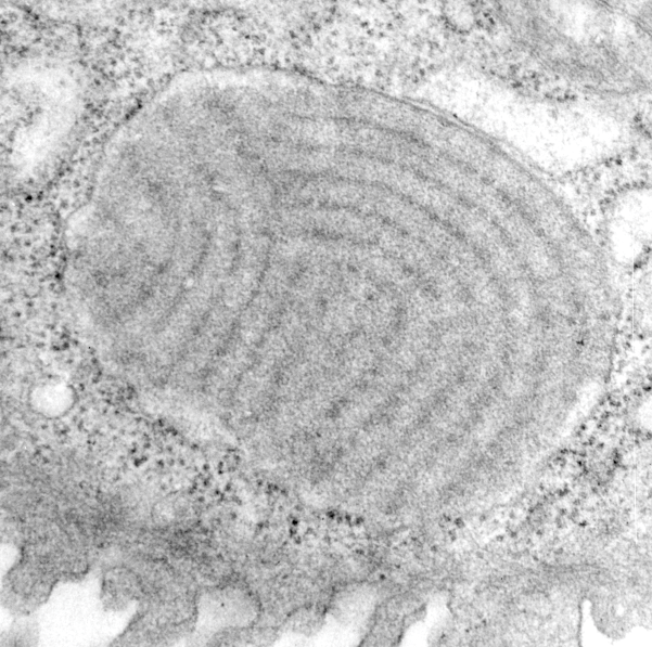
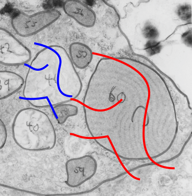
Negative 9879 block 23494 ferret # 16, untreated. Electron micrograph of an alveolar type II cell with a surfactant protein granule (which in some ways is similar to other collectin, c-type lectin, style granules, where organization of these proteins is highly oligomerized–e,g, Birbeck granules).  Embossed upward, and down ward images below. Bottom image is a cropped and enlarged from middle image, showing vertical lines which may be 4 or 5 per 100 nm and can transcend all three periods of this granule; ribosomes (as a size marker of @27 nm); periodicity of the medium dense layer of this granule (blue dots and 100 nm bar marker).

quintessential_SP_granule_shadowed-1quintessential_SP_granule_shadowed-2vertical-banding


Alveolar type II cell granule in ferret – electron microscopy

More on the periodicity of the dense outer bands of the alveolar type II cell surfactant granule.  Still think this is probably surfactant protein A. I have been assessing the densities in the darker outer bands, which are rounded molecular complexes, much more clearly seen on the tangential plane (yesterday’s images), and which looks to be something over 30 nm in diameter (using an adjacent ribosome as the size reference), and only two (with space between) appear in any 100 nm stretch.  The punctate nature of the lighter bands in this granule seemed to be more like 5-7 per 100 nm, so there is an obvious difference, both in size and in spacing of these two entities. How do they knit together into the granule?.

Dense bands are more continuous than dotted, particularly views perpendicular to the length of banding, but I would suspect that differences in the appearance of dense band periodicity has to do with the way the proteins denature depending upon the constituents of the fixation fluid, whether more or less calcium is present, or whether it is more hypertonic, or isoosmolar, but this is conjecture.

Figure below shows the intermittent densities in a perpendicular cut, unlike the post yesterday which showed the dense band densities on a tangential cut (and more pronounced). Figures below are arranged thus:

  1. quintessential alveolar type II cell surfactant protein granule UNRETOUCHED- Electron micrograph shows a granule with three linear periods and a ballooned end. Diameter of one ribosome is taken to be 27 nm, one period 100 nm (vertical black bar marker) therefore the whole granule here is approximately 300 nm in height (thickness). TOP
  2. quintessential alveolar type II cell surfacant protein granule with ribosomes which are on growing ends (and in this particular image, on the balloon end, and a couple of ribosomes on the opposite end, with membrane without ribosomes parallel to the long axis of banding. Ribosomes are PINK – and – the
  3. quintessential alveolar type II cell surfactant protein granule itself, is BLUE
  4. measurements of periodicity in dense as well as light bands BOTTOM, with green line over green dots (representing about 15 nm diameter punctate periodicity for lighter bands and red line over red dots (30+ nm diameter) showing periodicity of dense band protein molecules.

quintessential_SP_granulequintessential_SP_granule_overlay-1quintessential_SP_granule_overlay-2

Alveolar type II cell layered granule in ferret: even the dense bands have periodicity

Alveolar type II cell layered granule in ferret: even the dense bands have periodicity. I had generally thought that the outer dense bands of the layering in alveolar type II cell granules of the ferret were pretty continuous. It was easily evident that the middle band in each period of the multilayered granules displayed a periodicity (5-7 per 100 nm), but I had not seen much periodicity in the more dense outer layers of each period.  On several tangential sections of these granules, in the ferret, it seemed as though there is also a periodicity in the tangentially viewed dense bands.

It seems that if one measures a nearby ribosome at 27 nm, that the the distance between bands here (spread just slightly because of tangential sectioning) is just greater than 100 nm, and that the darker outside bands have areas (highlighted in pink using photoshop eraser and burn tools and overlaid on the original micrograph (top diagram – dust and scratches removed without changing data) then the distance between the center of each enhanced rounded periodic dense area is about 100 nm from the next.

6711_23303_ferret_136711_23303_ferret_13_aAs with almost all the granules seen in this study, the above shows ribosomes where there is addition of new protein to the granule (left and bottom parts of the granule above) and the area where the granule is NOT growing, upper right, the limiting membrane is ribosome-free.

This particular granule is pretty close to the apical plasmalemma (i should have oriented these cells with the microvillar surface “up” probably, but this distance is typical in my opinion, no granules have been seen exiting the plasmalemma (not apical, basal, or lateral) – so this raises some issues.

Alveolar type II cell intra-RER protein organization: ferret, more hexagonal structures

Alveolar type II cell intra-RER protein organization: ferret, more hexagonal structures per the last two posts, there is more order here,  actual linear densities perpendicular to the long axis of the layered granule, and these blend out into the tangential plane as hexagonal structures with a central dense area (either 1 or 2 dots — in this case mostly 1 dot).

Top electron microgr9855_23349_ferret_17aph (unretouched, except to show the bounding box of the inset image) is an enlargement from a negative (9855) of a ferret type II cell, of just one of the layered granules present which is particularly well suited to examination of its tangential-orientation and detection of patterns that might indicate that it is surfactant protein A.

Bottom electron micrograph is inset, enlarged, and the patterning perpendicular to the long axis of the granule layering (here, tangentially cut) is seen, and the erase tool has been used to outline what could be hexagonal patterns for the surfactant protein A 18-mer bouquet.  You are free to decide if this looks right. My question is that they are small, but also would represent just 1/4 of the 100 nm width of the real pattern-period, of mirrored and stacked (4 molecules) of SP-A 18-mers. I have not highlighted all the vertical lines, nor have i highlighted all the hexagonal structures.

9855_23349_ferret_17-inset


Alveolar type II cell intra-RER protein organization: ferret

hexagonal_proteins_in_alveolar_type_II_cellsMore tangential sections of RER layered protein from ferret alveolar type II cells seen with electron microscopy. I have chosen one of the more easily identifiable granules with the light portion of the banding ‘periods’ spread out so that any order seen on from the “top down” might be highlighted.  I have outlined in red (this is a “partial erase” in photoshop, against a red (and green) layer), no other manipulation of the micrograph has occurred.

The red outlines are pretty obvious hexagonal structures, seemingly mostly with a central dense area, but way to often for chance, two central dots.  I have seen this so many times that it becomes necessary to call attention to it, and likely it means something in the organization of this surfactant protein (which I am calling surfactant protein A). On the other hand, the size of a ribosomes in these micrographs would make the hexagons a little too small….  maybe only 75- 80 nm across, so this is an issue to reconcile.

Red outlines, within the tangentially spread inbetween layers), green outlines, an area which might be still within this particular RER profile, but might actually be cytoplasmic.  I have compared the incidence of cytoplasmic heaxagonal structures, there are some, no question, and I don’t want to read more into the hexagonal molecular organization than is warranted.  To me however, it is significantly greater within the membrane of the RER when tangentially sectioned as happened here, and the previous post (same animal, same micrograph, same magnification, different site.


Factors that may influence granule layering patterns in alveolar type II cells

The granule in the alveolar type II cell of guinea pig, ferret and dog is unique, in that it appears to have wonderful shapes, concentric, U-shaped, linear, branching (even highly branching mostly at perpendicular planes to the original layering). I was trying to figure out what could cause such variation in shape. One keeps in mind that these are cross-sectional images, 2D of the 3D cytoplasmic granules, and therefore can be difficult to interpret as a whole granule.

To note first: there are often parallel “jets” (which I have called periods, just to be proper ) for the production of surfactant protein A (yep, I am calling it that with nothing but circumstantial evidence). Sometimes there are 10 or 15 such nidi of production, each perpendicular to the long axis of the layering (banding) and having about 4 ribosomes (the 4th ribosome in each period serves as the 1st in the adjacent layer).  The amount of resistance at the opposite end of the granule to protein synthesis, and the rate of protein synthesis (at the growing end(s) of the granule where the ribosomes are ) will determine whether this “jet” will spew out SP-A in an unhindered stream to create a linear granule or whether it will buckle, twist, spin or curl.

I began searching extrusion images on google, and these below (easily-found set of 6   extrusion images which illustrate the point) do a really good job of matching what is seen electron microscopically, of what becomes of the structure of protein layering during SP-A production when the protein produced meets with resistance.  Awesome… ha ha, a natural physical phenomenon, I will bet. What scientific journal will publish my diagram… ha ha, probably not.  Too funny.

extruded_soft_serve_polyethylene_pasta_model

Alveolar type II cell, hexagonal order in granule tangential sections

Comparisons between granule and adjacent cytoplasmlooking for hexagonal or bouquet structures in the alveolar type II cell RER profiles that might be a surfactant protein, such as SP-A. Image below has two portions of cytoplasm outlined (same size), one at tangential areas of a cisternal body – aka – surfactant protein granule in the RER of a type II cell, and the other in nearby cytoplasm.  The redish tinge is created with the eraser tool in photoshop, where I added transparency around areas which appeared to be hexagonal structures with a central dot.  The central dot seems to be an important morphological portion of these hexagons.  See the cytoplasmic rectangle (as opposed to the rectangle from within the granule) to compare whether this might be random, artifact, or possible a reflection of the fixation (yes it is artifact, tell me something new) changes in this protein (I am calling surfactant protein A). There is a difference, I have not made an extensive comparison between intra granule, and extra granule cytoplasm and the general occurrence of 20-40 nm hexagonal structures.  Someone could certainly do that. It is a granule from ferret 9855_23349_#17). Judge size by ribosomes (@27nm) see the red outlines of prominent hexagons in the square within the cisternal body (granule) and count them again in the rectangle over cytoplasm (also enhanced with red as mentioned above).

hexagonal_bouquets_type_II_cell_granules

Alveolar type II cell RER granule and adjacent lamellar body

Alveolar type II cell RER granule and adjacent lamellar body from a ferret, in this case untreated, a block from peripheral lung makes a pretty interesting statement. Not withstanding the bad electron micrograph, and my writing from 35 years ago, at the very moment of encountering this image I was wondering to myself, and creating a spreadsheet, about the impetus for this protein to form circular, and linear, and u-shaped patterns within the RER profile, and side by side, was a lamellar body which had similar configuration. Yes, fixation is not what could be seen in today’s labs, but it was what was recommended in the 1980s, but the similarity of the lines is pretty much an indication of something besides chance.  In retrospect, the same variations seen in sections of lamellar bodies are seen in these intracisternal proteins: linear exterior, mostly linear protein banding, round exterior (just like lamellar bodies) concentric banding), U-shaped exterior, semi-curved banding. some times banding branching, or forming x or y shapes. It is not difficult to suspect that some membrane forces are at work in the formation of these similar shapes. Not my area of expertise, but certainly obvious here.  This image is neg 6598_block_23139_ferret #11 untreated.  Red lines flow across the pattern of layering in the alveolar type II cell granule, and blue lines flow with the direction of lamellae in the lamellar body adjacent to it. Grey outlines are left over from morphometric studies for a different purpose (sorry about those, but I didn’t take time to rub them off). If you need a measure…. the layers, or bands in the granule (labelled C) are just over 100 nm because the section is tangential, otherwise bands would be at 100 nm.

6598_23139_ferret_LB_CB