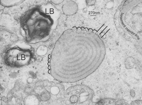
Electron micrograph of a guinea pig alveolar type II cell which shows a granule of layered protein in the RER, which has a concentric arrangement (almost like a braided rug – for those of you who know what these are). Along the periphery of the granule however, there are at least two areas, maybe three, where the layering (banding) has branched out perpendicularly to the concentric or wrapped original granule. black lines indicate the direction of new layering nidi, and the scalloped lin shows areas which are lilely to be the marking points of each “period” bar marker is 270 nm based on the dimensions of a ribosome from this same micrograph. 96813_17084_guinea_pig_301 (untreated).