I am getting bored with surfactant protein A: Taking a break to look at some old micrographs from mice that were infused with FC47 (FC standing for fluorochemical) emulsions. This particular mouse (No. 8) was given 75cc/kg of an emulsion (10% in 5% F68 – emulsion number 741028) on four consecutive days (10-28-74 to 10-31-1974). The mouse weighed 22.2g at sacrifice (11-1-1974), liver =1.79 g, spleen=0.62g. Tissues were fixed in Karnovskys, cacodylate buffer 2% OsO4, dehydrated in ethanols, embedded in Epon 812.
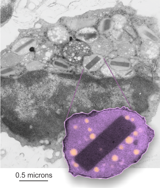
The unretouched image is grey scale in the background, one granule with perfluorochemical “droplet-footprints” remain in this (and many of the other) granules in this eosinophil. Components of the granules include histamines, peroxidase, ribonuclease (RNase), deoxyribonucleases (DNase), lipase, plasminogen, and major basic protein (stored in the dark crystalloid like structures within granules). Inset is contrast enhanced, cut out, enlarged, FC47 droplets pseudocolored.
The propensity for the minute emulsion particles phagocytosed by eosinophils to end up in the granules is something interesting in and of itself. Several types of granules hold the little PFC (perfluorocarbon) footprints, not just those with MBP crystalloids.