Alveolar type II cell intra-RER protein organization: ferret, more hexagonal structures per the last two posts, there is more order here, actual linear densities perpendicular to the long axis of the layered granule, and these blend out into the tangential plane as hexagonal structures with a central dense area (either 1 or 2 dots — in this case mostly 1 dot).
Top electron microgr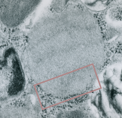 aph (unretouched, except to show the bounding box of the inset image) is an enlargement from a negative (9855) of a ferret type II cell, of just one of the layered granules present which is particularly well suited to examination of its tangential-orientation and detection of patterns that might indicate that it is surfactant protein A.
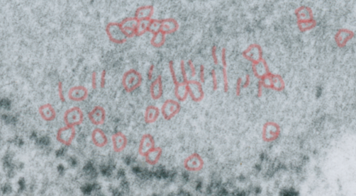
Bottom electron micrograph is inset, enlarged, and the patterning perpendicular to the long axis of the granule layering (here, tangentially cut) is seen, and the erase tool has been used to outline what could be hexagonal patterns for the surfactant protein A 18-mer bouquet. You are free to decide if this looks right. My question is that they are small, but also would represent just 1/4 of the 100 nm width of the real pattern-period, of mirrored and stacked (4 molecules) of SP-A 18-mers. I have not highlighted all the vertical lines, nor have i highlighted all the hexagonal structures.