Monthly Archives: November 2016

Rabbit alveolar type II cell: criteria met for protein granule?

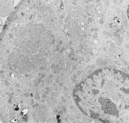
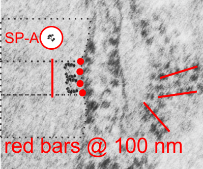
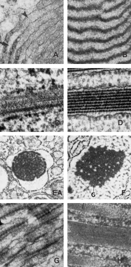
I revisited the protein acumulation in the alveolar type II cell of a rabbit (this particular rabbit was 79 days post infusion of an emulsion of oxydecalin 10% and F68 25% at 60cc/kg intravenously, on 9 9 1987.  negative 11320, block 31750, and this is one of only two locations where there was a stacked (or single) profile of RER which had some of the characteristics of an intracisternal protein that might be made up of surfactant protein, in particular surfactant protein A.  The criteria met include: 1) stiffness of the portion of the RER which has the protein inclusion visible, 2) loss of ribosomes on the longitudinal axes, but ribosomes on the growing end(s), 3) some evidence of layering within the granule (see inset where i highlighted what i think is evidence of layering) 4) a parallel stacking. I have considered this an equivocal demonstration of a surfactant protein granule in the alveolar type II cell, since the layering is really not that prominent.  The bigger issue is that, if one considers the ribosome of the rabbit, to be similar in size to other mammalian ribosomes (I have taken as an average about 27 nm), then the width of the profile at its narrowest dimension is about 1.5 times wider than the layered granules seen in guinea pig, ferret and dog.  So this is an issue.  See for yourself in the image below. (two prominent scratches stamped out using photoshop, contrast increased also, otherwise no processing). Red dots=ribosomes, dotted boxes show areas of inset. Insets show areas of possible banding which I highlighted with the burn tool in photoshop.  So original on right, emphasized on left.

rabbit_alveolar_type_II_granule_SP-A
While you are on this page, check out the awesome glycocalyx on the apical plasmalemma folds and projections in the upper right corner of the electron micrograph.

Hamster: alveolar type II cell

Another example of a species in which I have hunted from layered granules in the alveolar type II cells is hamster (though only a few animals have been studies). There are, as in cat and rabbit) frequent examples of RER which has a slightly dilated appearance, and also has obvious protein density within the lumen of the profiles, but lacks all but the very faintest appearance of layering, and also does not abide by the criterion for layered granules in that ribosomes stud all aspects of these RER profiles, and the rigid looking, ribosome free, part of the granules seen in ferret, guinea pig and dog, thus one has to assume either that surfactant protein A is sufficiently unique in the three species above where granules can organized into a regular substructure, or that something else altogether is responsible.  hamster, male, animal# 3, neg 7278, block 24733,

hamster_lung_type_II_cell

Cat lung: alveolar type II cell

Cat lung: alveolar type II cell, with arrows to three profiles of endoplasmic reticulum which have a structure not typically found in alveolar type II cells of common laboratory species (mouse, rat). These are rounded and it is not actually that hard to see some striations in them, particularly the one on the most left.  In a series of micrographs from untreated cats (taken incidentally from other studies) no sturctures with highly visible layering in the RER were seen, which doesn’t mean these profiles are not surfactant proteins, it just means that they are not organized as seen in ferret guinea pig and dog alveolar type Ii cells in other studies. negative 7268, 24564, cat #3, untreated (gender?, age?)

cat_lung_type_II_cell

Rabbit lung: post intratracheal instillation of iron oxide and dibenzo cabazole

Rabbit lung: post intratracheal instillation of iron oxide and dibenzo cabazole, electron micrograph. This is not a great picture, but i post it just so one can appreciate that there an area of odd membrane swirling in the middle left, and a small area just like it in lower left. The cell has a lobular nucleus and there are iron oxide (probably with DBA alongside) within this cell. I don’t think it is an alveolar parenchymal cell, but likely an inflammatory cell. A type II cells from the same microgrpah has lamellar bodies, which this cell does not, which further indicates it as a migratory cell. Animals given DBC do get tumors. neg 4447, block 17793, Rabbit #32-3.  Sorry the micrograph has stain ppt on it, but it is worth looking at just for the layered structure.

rabbit_lung_iron_oxide_dibenzo_carbazole


Kroger doesn’t listen: potential food hazard – faulty frozen food packaging

I really don’t like it much when I go to a manager of a grocery store and explain that they are selling a product that has the potential to be contaminated, and they don’t pay attention. Kroger would be such a store. It has been easily a year since I first mentioned to them that their packaging on some frozen fruits (my experience has been with Private Selection) in that it has the potential to allow bacteria and other non-food things to get inside the packaging.  I first noticed the packaging issue with their dark cherries (frozen) and when my freezer went through its defrost cycle, the liquid inside the packaging leaked out of the package and onto the bottom of my freezer.  This in and of itself is not the issue, though I did have to clean up a mess.  The greater issue is “if something can lkroger_doesnt_listeneak out” of packaging, then “something can get in”…. that makes sense to me, am I the only one that it makes sense to?  So I called their customer service hotline, and explained the “potential hazard” and expected a response.  But none came.

I made several other calls to them with different packages of frozen fruit, the calls in the middle of the summer were about frozen blackberries.  So this last time I got a package (this time a small package) of frozen blackberries I noticed that they had not heeded the information sent, and that they are still packaging their food with a potential for contamination.  Below you will see the perforations in the packaging at the top, presumably to make it easy to tear open (which I doesn’t anyway) and those perforations are relatively large holes, big enough for insects, big enough for fluids from other sources to seep in.  Why is a company so deaf to potential litigation on product contamination?  The reason I would propose is that they think they are too big to fail.  A common theme in todays world (which unfortunately even spilled over into politics this presidential election year) .  Call your Kroger company and complain if you eat these frozen products, you might save yourself, or someone else from food-borne illness.

THe white dots on the lower picture is “light” coming in through the holes in the packaging. The package’s zip loc closure is NOT sealed when the product is produced thus all during production, shipping, storage and to your home, it is open for contamination.


4 ribosomes – four SP-A molecules?

Another view of an image of an RER granule from an alveolar type II cell. where an actual SP-A shadow cast molecule (from the internet) was duplicated, mirrored vertically, both copied again, and pasted so that four molecules are stacked. These then were placed next to an opportune profile of an RER granule which clearly showes 4 ribosomes in a period….  As previously mentioned, one ribosome for each of the most dense layers (that is at the widths of the single periods) and two ribosomes within the period, one on either side of the central dense band.  4 ribosomes per period in all….  which pretty much matches the concept of four SP-A molecules end on end as imaged here. Still thinking on this, comments (to my personal email, not on this blog page) are welcome.

ribosomes_SP-A-4_and_4

Regular protein layering in RER

I was looking in the literature for examples of layering or high organization of proteins within the RER of various cells. There is no dearth of examples. Many really wonderful photographs are found in the book edited by Feroze N. Ghadially.  Ultrastructural Pathology of the Cell and Matrix: A text and atlas of physiological and pathological alterations in the fine structure of cellular and extracellular components, Edition  3, Volume 1, Butterworths, London, 1988  THESE IMAGES ARE FROM THAT BOOK AS AVAILABLE ONLINE, JUST SCREEN PRINTS AND CROPS (without permission btw).

inclusions_in_RER_layeredThere are some very awesome pictures which show enormous variation in the orderly arrangement of proteins (mostly in pathological situations, as is likely for the alveolar type II cell that might be packed with surfactant protein A in the RER).  Some show some of the characteristics of become ordered at the transition from RER to golgi, sort of signifying that something has be modified in the protein, or the RER microenvironment that has allowed for alignment of the protein molecules. A, banding with heavy lines and very precise spacing B, bands or layers of very different densities – and also widths C, tubular appearance C, D, central aggregagion spacing after protein synthesis, abundant ribosomes on the whole granule (unlike the alveolar type II cell granules where there is a definite directionality) E, F, parallel and perpendicular directions G, and numerous layers H.

Moons at perigee

With all the hoopla about the close moon Nov 14 2016 I decided to really try to see if there was consensus on the ‘man in the moon” which i see differently apparently than others.  In the east i see a smiley face man, squinting left eye, and a long nose and half open mouth, and in the setting moon, there is a sad face with a downturned mouth (former left eye) and a tiny circular nose.  The rotation of the face is about 30 i think.  Some astronomemaninthemoonr out there would know.  I resurrected two classic images from the “long ago archives of moon faces” and made this quick video with my own “man in the moon”.

LINKED HERE