Comparisons between granule and adjacent cytoplasmlooking for hexagonal or bouquet structures in the alveolar type II cell RER profiles that might be a surfactant protein, such as SP-A. Image below has two portions of cytoplasm outlined (same size), one at tangential areas of a cisternal body – aka – surfactant protein granule in the RER of a type II cell, and the other in nearby cytoplasm. The redish tinge is created with the eraser tool in photoshop, where I added transparency around areas which appeared to be hexagonal structures with a central dot. The central dot seems to be an important morphological portion of these hexagons. See the cytoplasmic rectangle (as opposed to the rectangle from within the granule) to compare whether this might be random, artifact, or possible a reflection of the fixation (yes it is artifact, tell me something new) changes in this protein (I am calling surfactant protein A). There is a difference, I have not made an extensive comparison between intra granule, and extra granule cytoplasm and the general occurrence of 20-40 nm hexagonal structures. Someone could certainly do that. It is a granule from ferret 9855_23349_#17). Judge size by ribosomes (@27nm) see the red outlines of prominent hexagons in the square within the cisternal body (granule) and count them again in the rectangle over cytoplasm (also enhanced with red as mentioned above).
Daily Archives: December 20, 2016
Alveolar type II cell RER granule and adjacent lamellar body
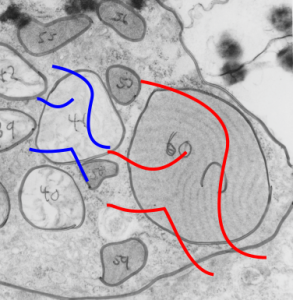
Alveolar type II cell RER granule and adjacent lamellar body from a ferret, in this case untreated, a block from peripheral lung makes a pretty interesting statement. Not withstanding the bad electron micrograph, and my writing from 35 years ago, at the very moment of encountering this image I was wondering to myself, and creating a spreadsheet, about the impetus for this protein to form circular, and linear, and u-shaped patterns within the RER profile, and side by side, was a lamellar body which had similar configuration. Yes, fixation is not what could be seen in today’s labs, but it was what was recommended in the 1980s, but the similarity of the lines is pretty much an indication of something besides chance. In retrospect, the same variations seen in sections of lamellar bodies are seen in these intracisternal proteins: linear exterior, mostly linear protein banding, round exterior (just like lamellar bodies) concentric banding), U-shaped exterior, semi-curved banding. some times banding branching, or forming x or y shapes. It is not difficult to suspect that some membrane forces are at work in the formation of these similar shapes. Not my area of expertise, but certainly obvious here. This image is neg 6598_block_23139_ferret #11 untreated. Red lines flow across the pattern of layering in the alveolar type II cell granule, and blue lines flow with the direction of lamellae in the lamellar body adjacent to it. Grey outlines are left over from morphometric studies for a different purpose (sorry about those, but I didn’t take time to rub them off). If you need a measure…. the layers, or bands in the granule (labelled C) are just over 100 nm because the section is tangential, otherwise bands would be at 100 nm.

