Please feel free to use any and all micrographs from this post. I did not invent the microscope, nor the techniques which are utilized, nor the equipment to section, nor the stains, nor the animals used in this research. To do as i see some (particularly this one dude, Dr. Jastrow whomever, egoist) who somehow thinks that he is responsible for every image in microscopy and watermarks these images like a tiny child as if — by obtuse correlation — he owns his own DNA sequence — or the images of animals and or plants he never created and only views because of countless inventions by other people. This is just an ego out of control and fearful. Makes me laugh and feel sorry for someone who has to protect what he neither invented nor understands. Sorry state, does he not think that he is standing on the backs of countless hard working scientists, and also trampling under food thousands of dollars paid for by external funding (in all likelihood). All this, and then collect revenue from google adds…. where is the justice in taking public funds and then publishing them for profit.
Daily Archives: February 1, 2017
Does alveolar type II cell granule exit at the apical plasmalemma?
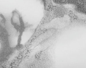
The highly ordered protein granule in the alveolar type II cell of some species doesn’t not often show any signs of where it “goes” in the cytoplasm, whether to exit at the apical plasmalemma or whether only to be dumped into the lamellar body to exit the apical plasmalemma with immature surfactant that the lamellar body holds. This is probably an image of the closest encounter I have seen between a granule and the apical membrane, and it is not complete, nor definitive at all, but just a reminder of the fact that the issue remains unresolved. See here four granules, the one on the left is very close to the apical membrane. There is a white line around a nearby apical membrane fold (typically called a microvillus, but certainly not a rigid one) and the bracket denotes the space between the former and the latter — close at best, but not a membrane fusion and release.
Banding is clearly evident within the granule. Sadly, the back of this micrograph is labeled “duplicate” but i cannot find the original with the description of negative number, block number animal number and species, but it will be either guinea pig or ferret.
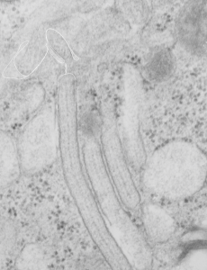 Just for comparison, here is a close encounter of granules with a lamellar body, which is actually a fairly frequent event. Lamellar bodies on left upper and lower right. three angled structures are granules (you can see the linear banding in the center one.