Monthly Archives: March 2017

University of Cincinnati: a new medical sciences building

University of Cincinnati: a new medical sciences building.  This really is sad. The medical sciences building built in the early 1970s is being replaced. I can only conclude the following:

  1. UC has too much money, thus can waste it
  2. UC builds a bad building that couldn’t last 40 years
  3. UC has new administration that has to “tag” the campus, just like a dog tags its territory.

none of the above is excusable. I was educated in the “first” college of medicine building, and watched the building of the “second” college of medicine, I wonder what mistakes will be made as the “third” college of medicine goes up.

Nuclear R-rings vs mokumu gane

I just love when science and physics come together. It makes me a believer in our abilities as human inventors and copiers. On the left is a black and white (as they always are unless someone screws with them in photoshop) transmission electron micrograph of a nucleus, and they have pointed out an R-ring which is an internalized portion of the ER from either just the inner nuclear membrane, or outer and inner nuclear membrane together. Kudos to them for their assessment (publication is linked above).  And kudos also to whomever fabricated this colorful (which it is unless one choose just black and whites) polymer clay mokumu gane piece. The latter is a common technique among clay-lovers, pushing and folding and invaginating colors into other spaces, that tries to achieve what nature has done, and does, all the time.  Fun!!


SP-A: three possible configurations to match electron microscopy of alveolar type II cell granules

Still working on this granule in alveolar type II cells. I think there are three levels of organization to the cylindrical and circumferential manifestations of whatever surfactant protein comprises these granules. The original 18-mer, well confirmed by numerous other scientists, on the left, relative to the size of a ribosome, and also with the hexagonal order seen in tangential sections of the outer dense layer of the granule. Middle image is the proposed SP-A fuzzy ball which seems to be a spherical organization of the same 18-mer, just with something different happening in the center region of the sphere. I can’t even guess what that organization (missing perhaps some of the CRD regions of SP-A?) to make things fit. This image also relative to the size of a single ribosome (red dot). Then on the right hand side is a double-layered cylindrical granule (this one likely from guinea pig) which has a diameter of about 200 nm, which means is likely has a column of four molecules for each radius. Size of the actual superimposed cylindrical period, and then a relatively sized – diagram…  sized relative to the other spherical or concentric configurations of SP-A.   The micrographs do pretty much dictate the arrangements, or at least that was what dictated the arrangements of the SP-A stick figure I used (which was vectorized from models of SP-A in publications.

Unconditional love : Unconditional trust

Unconditional love does not equal unconditional trust

I can love with unconditional love, but not trust that person. I have experienced it.  I am not sure which “love” can survive the lack of trust, which love of the several meanings that the Greeks have named.  Phileo, yes, Agape, yes, Eros, yes, Ludus, yes, Pragma, probably, and maybe even the self love.  So I can love unconditionally but be aware of deceit.

A busy cytoplasmic network in vivo: isolated, little blobs

Mitochondria are great little organelles. Looking over some archival electron micrographs, isolated mitochondria from hepatocyte with a gradual post-natal knockout of a gene critical to glutathione synthesis (details) can be found in Y. Chen’s publication. So I am looking for changes in mitochondrial shape in isolated preps that might relate to oxidative stress, but confess that I am also on the hunt for mtDNA, and anything that would resemble a  type of mtDNA protein complex, like the nucleoid present in yeast mitochondria. Nothing in this micrograph but I did find a curious pattern of small densities which I have boxed and highlighted in the inset.

Here is a micrograph from isolate mitochondria in the hepatocyte specific Gclc ko mouse.  Post natal day 21, h/h + N-acetyl cysteine (NAC). Neg 18315, block 78177, 20,000x. Tiny black bar in original unretouched = about 25 nm, and also in the enlarged and dot-emphasized inset. I wonder what that organization might be, but not mtDNA alone, but possibly little complexes?

Approving of presidents

“Two months into his Presidency, Gallup has Mr. Trump’s approval rating at 39%. No doubt Mr. Trump considers that fake news, but if he doesn’t show more respect for the truth most Americans may conclude he’s a fake President.”  THIS IS A QUOTE from the Wall Street Journal.

I checked several sites for approval ratings during presidencies. I found a couple of nice charts. It seems that presidents get a boost after just taking office, in approval rating that is, then the decline starts… It seems more like a pattern, an inevitable grumpyness and disappointment that changes promised in campaigns are not delivered.

So Truman leaves office with widespread disapproval (lowest of all presidents from 1945 onward).  The polls that give marks as high as 59 % are few, and I have to think that if 59% is the top mark for any president over the last half century, then indeed, we are a divided nation.

Taking the above “sweet honeymoon” period info from published presidential approval ratings,  Trump will end up down around 5% just like Truman.

And then go to wikipedia and find totally different results…. In my book, results should be results, but apparently not.  Wikipedia indicates that all presidents are within a few points of each other….  What gives.  These graphs for Bush (top) Obama (middle) and Trump (bottom) show that Bush went out on a low note, Obama was pretty much even 
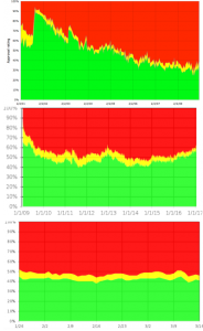
through the presidency….Trump (however short his tenure has been in three months…  Starting out low, will likely be no different than almost all president’s downward slide in approval till they leave office. As mentioned, this is an opinion pole, it may speak more to the polled and the pollsters than the president in question.  So much for Trump’s statements that we are all very “happy” with him.

The bottom line:  ha ha… write your own.

Inner nuclear membrane-outer nuclear membrane: inner mitochondrial membrane-outer mitochondrial membrane

Inner nuclear membrane-outer nuclear membrane: inner mitochondrial membrane-outer mitochondrial membrane, inner vesicular membrane and outer vesicular membrane, and when recognized there will be an inner Golgi membrane and an outer Golgi membrane, and an inner RER membrane and an outer RER membrane.  Just a matter of time before someone recognizes how to prove it.  Of course the first two are well established. It makes sense to have an RER which has this polarity.

And there you have it: totally useless abstract to a published paper

And there you have it: totally useless abstract to a published paper — and while I mean no disrespect for the authors, what are they saying.   And this is one of the journals I was considering to submit my manuscript on Surfactant protein A.
 
“A concise derivation of the principle of reciprocity applied to realistic transmission  electron microscopy setups is presented making use of the multislice formalism. The  equivalence of images acquired in conventional and scanning mode is thereby rigorously shown. The conditions for the applicability of the found reciprocity relations is discussed. Furthermore the positions of apertures in relation to the corresponding lenses are considered, a subject which scarcely has been addressed in previous publications.”
© 2016 Elsevier Ltd. All rights reserved.
They can have all the rights they want, who could read this and know what it said. ?? Heaven forbid that I ever wrote a meaningless abstract like this.
Ha ha, and what is more confusing is the summary…. which again doesn’t really say anything to me.

Isolated mitochondrion: electron microscopy

18327_78174_mouse#2 day 21 from_Glutamate-cysteine ligase catalytic subunit  h/h mouse given NAC. Fixation was in modified Karnovskys.  Isolated mitochondria. Condensed cristi. Some microsomes as well.  Typical mitochondria is about 200 nm in the smallest diameter. Original mag is likely to be about 40,000 (I will check) after 4 x magnification in enlargement. (which is useless, since i need a micron marker). Ref for specifics of the animal (h/h) are found in this article.