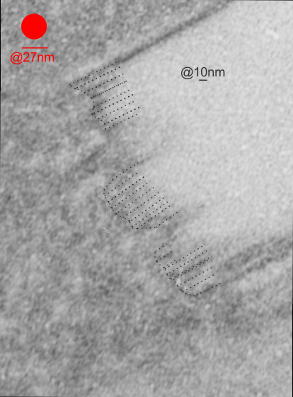
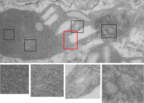
Two prominent areas of patterning are apparent in phagolysosomes in liver after IPFD infusion. The IPFD crystal itself, and at least four distinct protein patterns in the phagolysosomal proteins. The first image below is an enlargement of the red rectangle, and I have marked off with lines some of the pattern at the long end of the perfluorodecyl iodide crystals which seemed to work out to about 10nm in the short dimension. 23 measurements made at the end are marked off with dotted lines. A red dot=the dimension of a ribosome taken at the same magnification for reference. Below that is the low magnification image (unretouched — I removed a scratch from the top image but no data were altered as it was not in the crystal itself). Black boxes correspond to four very distinct protein organizational patterns within a single phagolysosomal body which also included the crystal in the top image. I am pretty confident that these patterns are a clue as to which lysosomes are present, and 4 may not be the only ones which show oligomerization into ultrastructural patterns. One thing to think about is that the kupffer cells (likely) in liver acting as macrophages which engulf (and hold onto for months) IPFD crystals will have a different panel of enzymes for lysosomes (and peroxisomes) than hepatocytes, and seemingly can become multinucleated under the conditions of massive IPFD inclusions.

