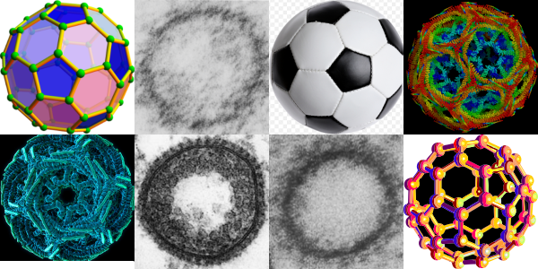
This is a wild goose chase looking for structures which explain and identify those which I have seen repeatedly in hepatocytes (and other cells), this time including clathrin coated vesicles and COPI and COPII vesicles. I am continually amazed how certain motifs repeat themselves in the microscopic, molecular and modern world. Stability in structure seems to transcend size. So image below is a collection of fullerene diagrams, some from my own electron micrographs, one from a widely republished electron micrograph (credits to the author if i knew who it was) and some from various wikipedia posts and of course the most widely recognized fullerene structure… the soccer ball. Just for fun but also tying in the concept that nature made it first. There is a difference in magnification from the upper left (i think around 1 nm – which actually sounds too big to me) — to the soccer ball 22 cm in diameter — well you can do the math 2,200,000,000 nm.
Daily Archives: August 15, 2018
Superballs vs fuzzy balls: immunity
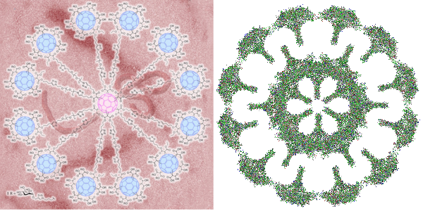
I got into searching for the structures of clathrin, COPI and COPII and one website visit after the other led me to this link. There headline was “Globular glycofullerene molecules prevent virus from evading immune system and entering cells” and I got goosebumps when i thought to myself that the surfactant protein A and surfactant protein D fuzzy balls might very well act as these proposed superballs in inhibiting infections from those viruses which have developed the reverse method for “entering” cells and avoiding the immune response (ebola and HIV for example). Seems a really fun thing to research. I posted a picture of what i designed as a surfactant protein A fuzzy ball (surfactant protein D makes fuzzy balls as well) and immediately saw the similarities between the octadecamer glycoprotein protruding in a sphere in both these fuzzy ball naturally occurring innate immune functioning proteins and the one that was produced by synthetically. Nature figure it out first.
BTW i dont like how the EBOLA virus is tangled within the backdrop of the globular glycofullerene on the left, called a “superball”, in fact it is kind of “nonesense” to me…. I could however envision the EBOLA virus stretched to oblivion as if stuck to a round velcro sphere, with areas exposed for digestion and removal. It has been stated that EBOLA type viruses (EBOLA is a “Filoviridae” virus and older than previously thought). That group of viruses has been interacting with mammals for several between 5 – 23 million years and it makes perfect sense that some sort of defense mechanisms have arisen in parallel. In fact a little bit of searching shows up this reference on filovirus entry.

 s
s