Monthly Archives: July 2018

Mitochondrial interactions with RER

Hepatocyte here, GCLC ko mouse, shows the donut and irregular configuration that is common in circumstances where mitochondria are stressed (I personally have seen it several times but in unrelated experimental circumstances so it is probably a generic response mostly).  In this really opportune section one can see a mitochondrion (with an odd donut shape) ont eh left, and a section of RER which has been sectioned tangentially showing the closeness and absolute regularity of the ribosomes along a spiral of mRNA.

I bet at some point all information about these association and the proteins (i saw a list that mentioned in the 800s and counting) in the inner and outer and cristae membranes, and the cristae junctions as well as the matrix will be modeled.  Until then, circumstantial evidence for the power of the interactions between mitochondria and other organelles (and cytoskeletal proteins) has to suffice.

The ribosome spirals here look to have approximately 7.3+/- .47 (SEM) ribosomes per spiral, n=9 (a small sample but the best orientation, and a single micrograph…. so this is just a suggested number obviously.  19735_73218_#201 liver alb+/- Gckc -/- postnatal day 28. liver mouse, no NAC.

One thing abou these mice that is pneumonic is the dilated ER, a mix of smooth and rough, ribosomes spaced and the presence of the little bubble-blip invaginations of ER within the outer RER membranes.  These mitochondria also display fission lines and tubular cristae, and quite a bit of it.  Blue dotted line (outline of one part of a mitochondrion – that one tangentially sectioned beside the RER, white box, area for enlargement to the right. Upper image on right has cytoplasmic ribosomes in mRNA-spirals (orange) and lower image is contrast enhanced to highlight the spirals of cytoplasmic ribosomes.  I really don’t think there are any good examples of mitoribosomes in either of the two mitochondria shown here.

Numerous proteins in the outer mitochondrial membrane (encoded by nuclear DNA) target and or are attached to cytoplasmic ribosomes. Cytoplasmic ribosomes have been visualized on mitochondria membranes (that would be the cytoplasmic face of the OMM). These are suggested to be linked by the translocase of the (outer) mitochondrial membrane (TOM) and it is reported that the ribosomes are in clusters (Till Klecker et al, 2014 Trends in Cell Biology) and that pretty much looks like these nRNA-ribosomal spirals… tidily wound.

MDVs, mitochondrially derived vesicles (about 70-150nm) are  purposeful buds of membrane derived from mitochondria which are targeted to lysosomes, and maybe other organelles.

Just an “aside” here, but is it possible that the donut shaped mitochondria (invaginations, and extra turns and indents) might be somehow a deliberate attempt to increase surface area for interaction with other membranes.   Another question is the orientation of cristae.  I have looked for cristae pores, and that relationship…  just not seeing it overtly.

Different effects from different types of trauma

I have casually logged into my mind what kinds of trauma to the skin turn into lichen planus lesions and which do not.  For starters, it seems that trauma from items which have antigenic properties (eg cat scratch, dog toenail scrapes, rubbing from a dog leash over the wrists) become real lichen planus lesions, where as more sterile injuries, bumps, pin pricks (as in sewing) dont have the same tendencies.  Another mystery is why “on me anyway” spider bites (1/2 inch red area with swelling and a central darker dot) don’t turn into lichen planus lesions.

I had a biopsy of a 30 or 40 year old lesion on my leg that was left over from small pox vaccination (which at that point in time was a conventional remedy for apthous ulcers, since it was believed there was some cross immunity achieved — that is not done any more, but because other herpes type viruses  can show post-infection increase in lichen planus i will be curious to see if that pathology matches others taken in my case.

Also noted, the impact of chemicals as instigators of lesions at points of trauma seems to be prevalent, eg, bandage adhesives.

While this disease requires the utmost of patience, and heroic efforts are required to keep from scratching, maybe there is healing somewhere for some people and at some point treatments will be found…. likely like one will have to say… I am a “recovering” lichen planus patient, as i don’t think the basic predispositions may be only genetic (as writen in genetic code or epigenetic modifications)  ha ha.  or not so ha ha but has an environmental (environment in the greatest sense that includes, chemicals, irritant compounds, man made compounds, diet, stress and the whole nine yards).  My sympathies to all you out there with this disorder. What is clear that diseases processes and problems that don’t threaten “life” get sent to the back burner in terms of research.

Mitochondrial proteins in membranes at desmosomal tethers

This concept of organellar interaction is of course old, and even has been on my mind for decades. I have made many posts on the tethers seen between mitochondria and desmosomes and hope at some point to understand and diagram the relevant collection of outer and inner mitochondrial membrane proteins (black lines above the membranes show area in question) present at the points of attachments to the intermediate filaments (red line shows direction) of desmosomal mitochondrial tethering. Box in top figure is rotated and enlarged for inset. Rat #9, liver 5983,  BH

Mathematics Visual – Matematicas Visuales — Geometric bodies

I was looking for help calculating the total height of a stained glass pattern that I was attempting to create for a diamond shaped pyramidal structure. I found it took me about 2 hours on google to even find the correct name for the structures in three dimensions.  This says nothing about google’s algorithms but it does say a lot about how little I remember about geometry (65 years past, maybe longer).

After looking over many sites I was totally overwhelmed with this one in particular and it is on a par with none other I have visited.  Check out the animation, the presentation, the whole site is really awesome.  However, I still have to ask for help — which I will do — for calculating the height of my pattern (and very likely, I wont find a single customer who would be able to enlarge it and calculate the height for themselves either… just to alleviate my own reduced self-worth at not being able to remember how to do it from 8th grade.

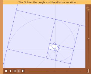
HERE is their webpage for an octagonal pyramid and for truncated pyramids. But looking further you will find many awesome pages, this one on the golden spiral.  Just peruse the site, you will be amazed. Here is a screen print from an animation.


LIFE IS SHORT — PRAY HARD

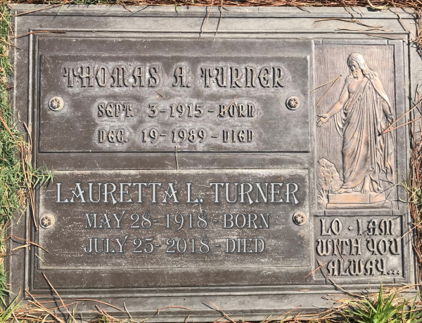
I was blessed to be born to Thomas Arthur Turner and Lauretta Louise Milner-Turner, in the the LA suburb of Eagle Rock, in the middle and post WWII period with 4 great siblings and a wonderful public school system, so much peace and friendliness. There could have been no luckier person than I.  That is not to say that life was perfect, no, but compared to the other 7,599,999,999 people in the world, I drew the luckiest card ever.  Thank you to my mom and dad and sibs and the world that was the 1940s to 1970s.  My mom will join my dad in a day or two. RIP


Retail spread and time-restricted eating habits

A really nice article on length of fasting time, and restricted eating times mentioned that there was a correlation with these and the levels of metabolic dseases common to obesity and overweight problems, diabetes, heart disease and the like. The studies were done by Hatori et al at UCSD using mice and various high fat and normal and ad lib and restricted feeding schedules but similar calorie inake.  The bottom line make good sense….. it is not only WHAT we eat, but really more about WHEN we eat and that relates to our circadian rhythms.  Second point here is the varying hours that many retail workers experience…. some early shifts, others late, and even those varying from day to day.  It seems pretty simple to me that this combination of “mall” food and changing “diurnal” eating patterns are likely related to “retail spread” of the waistline.

ImageJ

With all due respect to the writers of this image analysis program, it is so-not-intuitive. I have only played with if for an hour or so, but cannot even find where to click to delete a line…. This has to be so basic to a program, and also in accordance with standard keyboards…. as to be a careless? omission.
I have used lots of imaging software. I would really love to just know one thing…. how can i determine area using the line tool in CorelDRAW x15. I did not find that info on the internet, though newer versions may be able to do this. If so, CorelDRAW would be a great image analysis program. Can’t wait to see if further queries can produce instructions for using CorelDRAW for scientific imaging. It is a really nice vector program and with a few tweaks could be a great addition to the group of processing software packages. The selections and possibilities for analysis are way more sophisticated than those found in ImageJ. Wonder why CorelDRaw doesn’t come up with some kind of image analysis vector-type software.

Biological clocks : August lilies : Lycoris squamigera

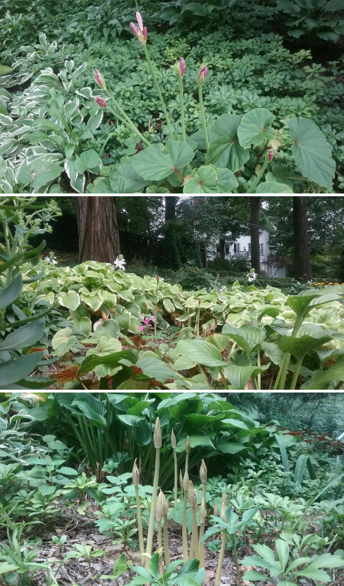
It was fun to predict when the august lilies would pop out of the ground this year. It was a pretty hot july here in cincinnati, and we had about 2 days that cooled off to the low 60s or maybe even the upper 50s at night. This made me think the bulbs would be on alert for the next rainy period. So after three days of rain, not terrible rain but steady moisture, i had 8 stalks of august lily appear from the ground, the next morning 32, the next morning (after a little more rain and cooler daytime and night time temps) i had more than 80. This afternoon (still cool but no more rain) there were more than 250 august lilies poking their heads out of the soil.

Biological clocks are amazing – (as an aside, one cover for JBC i did for someone working on biological clocks I have framed… he actually won a nobel prize for his work on bioloical clocks. I posted it here and also did an inside cover illustration (which i would have wished had been accepted as a cover here) but i dont think i have ever done much research on biological clocks in august lilies.

biological clock

Hardy members of the amaryllis family, emerge as astonishingly fast, with no foliage (which is early green and then dies back in spring) popping up from underneath hostas, begonias, ground covers etc. They also are known as Resurrection lilies, autumn amaryllis, august lilies, naked ladies, and as Lycoris squamigera, their botanical name.
Last year i dug up and replanted clumps (way over due…. decades over due) of 30 and 40 packed bulbs, so tight none could “breathe”. About three years ago i had over 200 blooms, then less and less and I didn’t know if it was the weather or that they were crowded. I did not anticipate that the new replants would bloom. Some certainly have.

I found this cute post from Stanford… Hacking the biological clock  . and a bulb society has a couple posts here.

FOR me….. it looks, year after year, it is a combination,  first cool nights, and lots of water.

and another post HERE that mentions the medicinal (toxic actually) compounds, and also this might explain why deer dont really like to eat my august amaryllis… or so i will call it from now on.

Earth temperatures

From my friend DWN.

Here is a list of natural causes that affect global atmospheric temperatures:

  • solar activity (frequency and strength of sun flares)
  • geothermal vents and underwater volcanoes cosmic ray flux
  • orbital eccentricity-axial tilt-and-precession of Earth’s orbit
  • magnetic effects of other planets
  • heat distribution between oceanic and atmospheric systems
  • changes in radioative forcing (balance between solar-radiation energy absorbed by Earth’s  surfaces and energy radiated back into space)
  • the large difference in land:sea ratio between the Northern and Southern Hemisphere

“Human activity” contributes something, but the effect is well under 1%, i.e. it’s not statistically detectable. (Yet. Could be in 100-500 years.)

Really nice video from NASA here.

Cristae

The numerous mitochodnrial variations (in total shape as well as cristae shape) seen with different species, tissues, strains, experimental manipulations can sometimes lead to interesting speculations about which mitochondrial membrane proteins might be present or absent or changed. Electron tomography as well as routine transmission electron microscopy can allow speculation about mitochondrial performance.

Isolated mitochondrial supply an easy example of changes in cristae junctions where under some conditions they are non-functional (missing).

In vivo, mitochondria are surrounded by areas of cytoplasm which can have cytoskeletal elements closely approximated, and/or linked to mitochondria (e,g, nerve terminals – called mitochondrial associated adherens complexes) , with desmosomes (sometimes called mitochondrial tethers–of which there are dozens of samples in many species on this website), nuclear pores (i don’t believe these have been named–of which there are also dozens of samples in many species on this website), and sometimes other organelles, such as RER and secretion granules).

Positions of cristae, arrangement  (perpendicular, parallel, etc) and number of cristae and cristae junctions are variable features depending upon which cytoplasmic elements are adjacent to that particular side (site) on the outer mitochondrial membrane. Some morphometric work was done on neural tissue by Perkins et al, Journal of Neuroscience January 20, 2010, 30(3):1015–1026, and they state that the number of cristae junctions is greater on the side of the mitochondrion which is involved in what they call “mitochondrion-associated adherens complex”  –maybe an unfortunate use of the word adherens…. owing to its immediate confusion with adherens junctions… maybe deliberate use of the word adherens because of the presence of what they term cytoskeletal structures).

estimates:

crista junction 15nm diameter
OMM thickness 20nm
omm+imm contact distance 14nm
tubular cristae (hepatochte mitochondria) 30-40nm diameter
about 90 degree angle (perpendicular) cristae to junctions
junctions at higher numberical density on sides where contact with plasmalemma is (specialized areas)
intermediate filaments and microtubules aligned with the long axis of mitochondrion (what does this mean? parallel?)
struts (perpendicular to mitochondrion, filaments a little thicker than intermediate filaments?) (a ring?)
filaments and microtubules define a “vesicle” free area as the M-associated adherens complex.
possible vesicle tethers beside the m-associated adherens complex about 8 nm apart