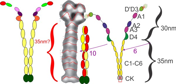
Lof et al, (Plos 1 Jan 2019) shows two monomers as a diagram of two molecules of von willebrand protein at a point where the winding portion is drawn apart. The actual AFM images look to me like the molecule winds up from the C term, and it makes sense in terms of ease of unraveling the molecule into the extended state at the beginning of clot formation.
Issues remain between diagram and AFM image: the diagram (even though the domains are named as C1 – C6) and puts six vertical ovals in the diagram, I also don’t see that number of densities (which plot on the LUT tables, in fact there are more, in addition to the clear appearance of two peaks in the LUT when lines for analysis are drawn horizontally on a vertically oriented image. Also, the top of the wound monomer in the AFM image is nothing like the diagram they show. I acknowledge that I might be missing something but the images look very different than the diagram (see their diagram on the right hand side) my analyses of LUT peaks in their image and measurements of theirs are certainly not the same proportions as the AFM image.
Here is a comparison image, my graphs on the right and my LUT on the image from their paper (they published four images which had the same one spot, two spots, one spot, two spots — patterning in the “stem”.
Other authors (Berriman et al, 2009 PNAS) uses electron tomography to make a great “…..lattice drawn on half the pattern (their Fig. 4B) which shows that the VWF tubules are helices with a repeat of 360 Å in which 13 subunits make three turns (about 4.3 subunits per turn).
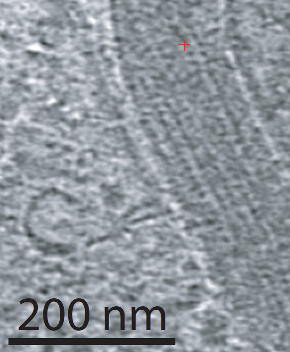
here is a great image of the granule (Wiebel-Palade body) in a publication by Berriman et al (PNAS) in which a very orderly form of tubules stores this protein. Great micrograph…. would love to do some organizing of molecules on this granule pix. ha ha.
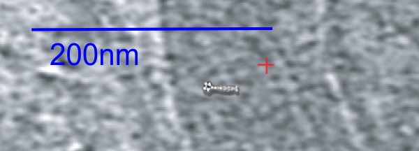
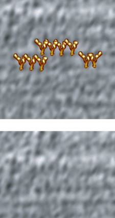
Dimensions of the shadowed molecule and the Weibel Palade body, don’t work out easily. but here is an approximate size highly wound up VWF protein over such a granule …within a granule, which can be a highly organized tubular structure….

This is just a wild hair…. looking at the periodicity in the electron micrograph and possible positions for a regular alignment of VWF in these Weibel Palade bodies…. Nothing scientificaly produced here just an image that looks right over an image that looks right…. ha ha, magnifications are not that far off… I think there would be vertical lines (as one sees in the Langerhans cell-birbec granules) were the VWF wound up at the C termimals (CK) to whatever binds them at D4. What is interesting is the alternating position of the densities along the margins of the tubules creating the illusion of the Y shape in the granule.


