Monthly Archives: March 2019

TOO LATE:

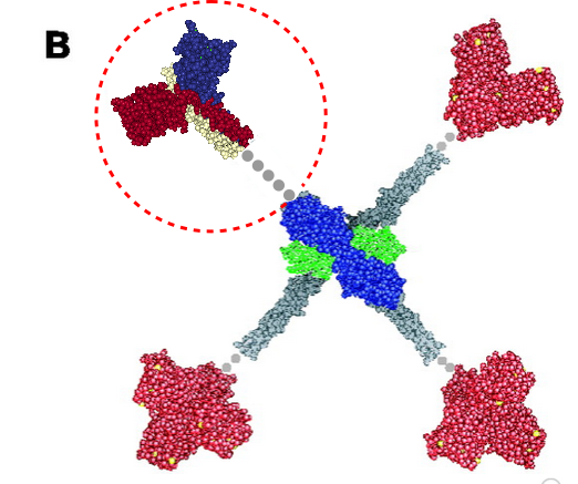
I get kind of sick to my stomach when i read that someone has discovered something that I found (recognized) a decade or more earlier, and know full well that there were people thinking of what i was thinking of even decades before that.Here is a cute article (i call it cute) because it was a worn-out thought in the early 1980s, and I had published at least two papers on the very close connections between mitocondria and cellular elements (desmosomes in particular) and also with nuclear pore filaments in a host of cell types (but mainly hepatocytes)…. Power to this woman, but shame on the editor for not searching back into the 50s and 60s where the wonder of higher resolution electron microscopy was burgeoning to finding out what the real masters thought (and even “moi” was thinking about the connection between organels and possible meaning). This is not to say that the isolation of the proteins and factors was known then, no, but the concepts were there.

What is silly is that I also pseudocolor and isolate organelles to play games with graphically… ha ha.  This site is full of examples. oh well.

Morphometry: SP-D dodecamers

I am hoping that some useful information will come from the development of this method of assessing AFM images, in particular here, of SP-D. The measures are actually 8 in number but i am not showing but debating whether it is necessary.  All dodecamers have been normalized to 100 nm arm length. Just one image (not my AFM but found in the literature) is shown, a test of the methodology.

Below there are measures in square nm for the carbohydrate domains (shown in the top part of the image), values for the angle of separation of the arms (only one acute angle is shown here, mean of the acute and obtuse angles for this image are given below); peak width is given, and peak height (both in nm) (from the deepest part of the valley on one side), nm2 for each peak (also from the deepest point on the peak), luminance is given (0-256) from the look up tables (obtained in ImageJ).  I understand that there are program s that can do some of these functions “automatically” but there are always details that machine vision really doesn’t “get right”.  This is an N of one, but this particular molecule was chosen (subjectively) as something very typical of AFM images purposefully in order to develop a technique.

So many things could easily be done…. for starters, comparing the nm2 of various CRD on the literally hundreds of CTLD (or even just the more well known c-type lectins, not to speak of arm arc angles and the position of the branching, and the N terminal connection width (in terms of nm).  All of the numbers below tell something about SP-D which need to be redone in dozens more images, and then compared to the variants of SP-D.

taking a good look at surfactant protein D dodecamers

I havn’t posted on lichen planus for many months:

I havn’t posted on lichen planus for many months: for you out there who have this (as I do), the years just go buy…and for me….stress is an instigator. best to you all.

I am studying “lectins” in general and know in my head somewhere that there is a connection between the lectins (involved in innate immunity) that i am looking at at any given moment might very well be critical) Little consolation from the itch.

I may have read this article before, but in todays search it seems like a good one.

Cant wait to see if the tooth that had a root canal (failed) and metal build up (now pulled out) will relieve my symptoms.  Hopefully.

Mannan binding lectin diagram: neck vs collagen-like region

More confusion in the literature surrounds diagrams (in my opinion) than text. Here is a diagram which I am not quite sure how to interpret, however, this publication states that the grey dots represent an as-yet-undefined collagen-like domain in mannan binding lectin (protein) (MBP 1HUP – MBL2, and 1KWT) and I have read countless times and tried to determine its structure myself for both SP-A and SP-D and not found anything of high confidence.  Yet the grey dots to infer “no yet determined” works for acknowledging that the molecular structure of the collagen like sequence in MBP (or the other C-type lectins) just hasn’t been worked out.. no that is not the problem, but it is that in my interpretation of this illustration, the grey dots are where the “coiled coil neck” is located NOT the collagen-like domain.  So in the RCSB, 1KWt) entry, (shown against the original diagram published by Teillet et al, 2005, I have pasted the image from the RCSB (which includes the high confidence structure of the CRD and the coiled coil neck) against the CRD and the “grey dot collagen like domain” they drew.  You can easily see my dilemma. My overlay is three color-for three chains in the trimer – of the CRD and neck, and grey dots for what I would have thought would be designated as the collagen-like domain, which then stops at the N terminus juncture (they show models of the serine proteases at the N terminus). The lower left and right and upper right arms of this cross-shaped mulecule have the dots (which they claim are collagen-like domains — where i think the coiled coil neck should have been drawn.

Variations in slicing and analyzing LUT tables: arms of an SP-D dodecamer

The best LUT plots for greyscale are not easy to obtain for curvy molecules.  This particular set of images and plots shows how the plot can be improved by some easy steps, none of which alter the data (the original image) but which produce cleaner results in terms of the LUT plot. (my name = #46 _SP-D, AFM from someone elses publication.)

Top image is the actual arm of the SP-D dodecamer just quickly isolated from the whole image using the eraser tool in CorelDRAW x5, and then cut into 10nm widths (vertical grid as shown), then ungrouped and centered horizontally and exported as a tif at 300ppi (next image down).  Getting the LUT plot using a rectangle did not show the peaks well, and a plot for a single line was obtained as well (with worse results). Since ImageJ does not have the potential to bend the lines at nodes, then some other technique for further straightening the area to be plotted helps.  The third image from the top is a manual vertical adjustment so the bright spots are aligned horizontally.  All those slices are groupd and recropped, and a grescale rectangle is placed behind with a medium greyscale color (125) to minimize the lines at each 10nm slice, and then it is exported as a tif (as above) and imported into ImageJ. THe resulting LUT plot is somewhat better. Number of peaks on each side of the N terminus is countable in both. In this particular set of two trimeric molecules the N terminus peak is not the brightest, but the leak on the left hand side (peak which would be the C1 portion of the collagen-like domain) (most medial peaks (right and left of the N terminus peak, the latter being central. The plots still seem to me to have three peaks in the collagen-like domain which might be born out on the left, not so much on the right half.


Here are two plots, red line is adjusted per bottom image, blue line is original (second image from top).

The other two trimeric arms are here (labeled c and d) were plotted with rectangle 500pxx@15px) blue plot line is a 1px line through center of the sliced and centered image. There is little difference. Three peaks between the CRD and the N terminus (which is in this case just a little to the right of center and only the tallest peak in the sampling of the rectangle) are found on the right. Three peaks are on the left as well, but they are grossly misshapen compared to the right hand plots.

SP_B: what shape

I admit to knowing nothing about SP-B, i am not going to shy away from commenting on diagrams that dont make visual sense to me however, and this is one such.  From a team of highly published and well recognized names in surfactant biology is a paper linked here which has a model suggested for SP-B. When i look at the hexameric original turquoise-blue ribbon diagram transparent behind their AFM image i am forced to ask myself…. why is a hexagon arrangement logical to them here when clearly the structure is a five sided polygon — at least in the context of this particular micrograph and diagram. This doesn’t make visual sense, no matter how much sense it makes to them scientifically.

Below. both background greyscale diagrams are theirs: on the left one i have superimposed a similarly colored regular hexagon pointing each of its angles to their blue ribbon molecular diagram which they propose to be SP-D (and to this particularly, i cannot comment )On the same image from their publication (copied and pasted on the right) i have drawn in a slightly irregular pentagon, which in my humble opinion makes more sense than their hexagon. (bottom image is  photoshopped to accentuate luminous areas. Still no real definition of a hexagon, certainly it is more complicated and nothing about the images suggests hexagon.

Nature loves copy and paste

Just looking at a few proteins on RCSB PDB lets us all know that for sure, unequivocally, without question, nature is great at copy – paste – mirror – rotate.  It is apparently an easy way to achieve variation. 1, 2, 3, 4, 5 and 6 (i am sure there are jillions more, i just stopped at 6). Great for defining new functions and structure-function relationships while conserving effort allowing for flexibility in evolution

C-type lectins: numerical summary

C-type lectins are an amazing group of molecules (well all molecules are amazing, LOL), part of the CTLD (C-type lectin-like domain) superfamily of glycoseaminoglycan-binding proteins: touted to be present in all multicellular animals, plants, and fungi and unicellular microorganisms such as a protists, bacteria, and archaea. C-type lectins function in glycoseaminoglycan synthesis, in enzymes, enzyme inhibitors, in coagulation and thrombin, and as esterases, dismutases and topoisomerases and growth factors.  They can be active inside or outside a cell; as cell adhesion molecules, as extracellular matrix proteins, as growth factors and morphogens, as lipid binding proteins, chemokines, lipases, annexins,

A-type lectins (someone will describe someday)

B-lectins (beta-prism lectins)


L-selectins (on lymphocytes —

MBL is a kind of prototype for this family of proteins. (working on this)

Calcium dependence was the key property of the compact carbohydrate recognition domains (CDR). But not all C-type lectins bind carbohydrates. It is clear that the use of the term CRD and c-type lectin domain are often subject to such broad use that confusion arises (review here  ).

Dimers

Trimers

First thing to clarify is whether the term “fold” which is used in many publications is really meant to be “the fold” or a more general description of a property, as in the “folding” plural.  I cannot see how “the fold” would be applicable since there are many folds in any one CRD.

Nice atomic force microscopy of mannan binding lectin

Nice atomic force microscopy of mannan binding lectin by Dong et al, though I could not use my method of measuring LUT plots on them. Nevertheless, in some of their images there was clearly hexagonal distribution and I have rearanged, marked and reposted some portions of their AFM image here and superimposed the carbohydrate recognition domain, at a slight angle (as would be projecting up from an N terminal (not shown) binding of six trimers.  Many many (almost all)  other researchers diagrams of show MBL as an oligomer with 6 trimers, not just three. The selected AFM images are “top down” the angle being 30o, the angle of the three diagrams is from the side.  I don’t know why the difference here. (top image by Dong et al, bottom image is from google images no authors listed in this post but they are readily found online).