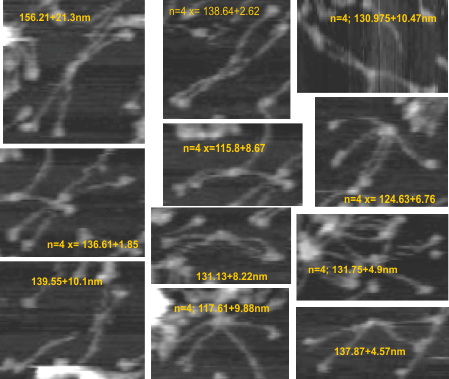
11 dodecamers were analyzed (some really hard to define in and amongst the various parts and pieces of SP-D in the multiple-molecule AFM image that is provided in a manuscript by Heartshorn et al on SP-D. Just 11, and four measurements using three different techniques were made to compare methods ultimately to get a gold standard for SP-D size from which to do brightness analyses of the collagen like segment of the molecules (where total length needs to be controlled (adjusted) to determine whether there are common peak heights in at least 4 consistent blips SP-D, two of which have been noted, the largest blip being called by Arryoy et al a glycosylation site.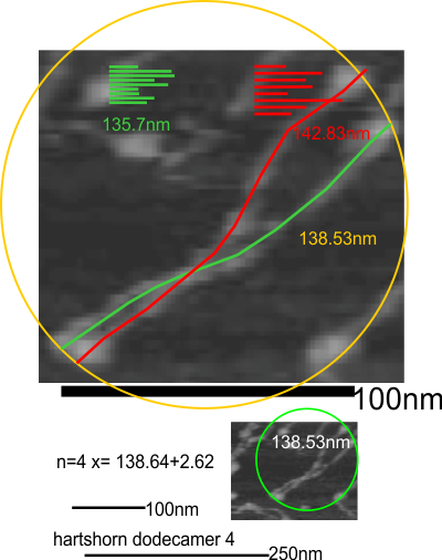
So here is a measure : diameter from original small image, diameters from images enlarged (remember that the diameter must lightly touch the outside of the CRD in three of the four CRD domains – one (two: one done on the original micrograph which is about 4 times smaller than each enlargement where diameter is also measured). Hartschorn et al’s bar marker for nm and peak height is used with all their image. Secondly, vector line measurements of each of the hexameric arms (alleged linking of two trimers into a hexamer at the N termini in the center of the molecule (with the measure of the hexamer going from one CRD through the joined Ntermini and exiting half the dodecamer with arms.
n=42;132.489+14.6