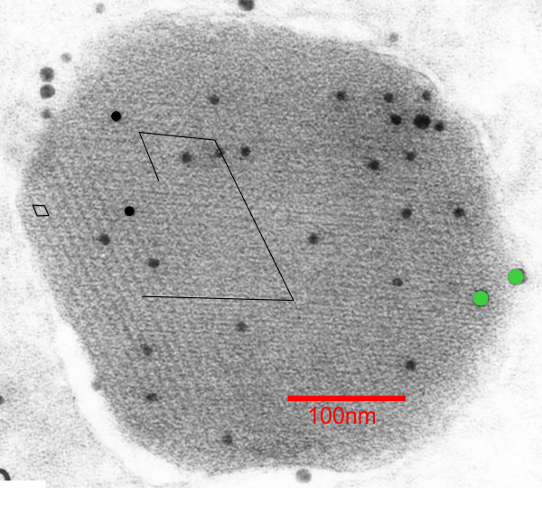
When I saw this picture (ref here) and saw the angles, rows and hexagonal array of bright and dark spots in this immunohistochemical image (SP-A they say is 10nm probe and SP-D is a 15nm probe and the SP-A is really the only one within the granule, the SP-D is on the peripehery and only two grains are present) I got curious to see whether there was any similarity in size of these “flowers” “bouquets” in a highly organized grid pattern and what I saw in the type II alveolar cells (posted dozens of times before now). Magnifications don’t really add up, but that is nothing new, since most magnification markers are way off what is actually reported for size.
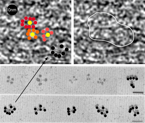
I think it is fair to say the probe size is pretty reliable (LOL). I put this image (with my colored (mostly 6 branched (presumably trimers of SP-A) flowers in color just to show them off, and marked two of the 60o patterns in a moderately enlarged image (sized with the colored flowers) against the backdrop of the clara cell granule.
The image of shadowed SP-A is one i have referenced many times, and the 50nm marker for those molecules would appear to be too large for the approx 13-20nm colored flower hexagonal figures within the clara cell granule however, the shape really fits – who to believe, perhaps totally irrelevant (but i think not). Images below, their granule. black circles=10nm, green circles (SP-D size)=15nm. Bottom picture, my depiction of the six sided SP-A in an hexagonal array, some CRD indicated by colored circles, N termini are central in the flowers. (someone elses’ shadowed image of SP-A 18-mers which sadly are not the right size..but i bet there is an explanation. White line in figure to right shows where in the cropped enlarged image i put the petals to the bouquet flower. 18-mers of SP-A I have drawn in lines and circles over their original images of shadowed SP-A molecules.