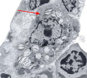
There is a book called Diagnostic Ultrastructural Pathology – A text atlas of case studies. Volume 1 ( Ann M. Dvorak, Rita A. Monahan-Earley) which on page 218 posts figure 182 (reproduced here – I thank them). It shows clearly that, while they were interested in lamellar body ultrastructure before and after ozone exposure, you can be sure that the most important structure for me is the “cisternal body” that flattened and layered protein structure that I see in the upper right-ish corner of their micrograph. Guinea pig must have some very robust mechamism for responding to environment, producing proteins (presumably some surfactant proteins) and in this case, after ozone they do not seem to become more prominent (according to this chapter in the book) as they do in dog (see previous post), but actually become less conspicuous.
Dimensions and position within the cell are classic “cisternal body” which I really wish I could rename SP-granule. Red arrow points to the RER granule.
