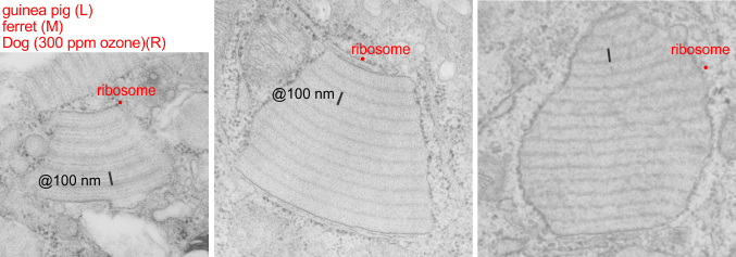
Electron micrographs of portions of alveolar type II cells: Two micrographs on the left are from my research, guinea pig on the left, ferret in the middle. The image on the right comes from a publication in 1973 by Stephens, Freeman, Stara and Coffin which had to do with exposure of beagle dogs to ozone. The claim that ozone increased these protein bodies (which for all intent and purposes match the protein body granules that I have seen in my animals), which also increased with age, particularly. I attribute these to some kind of immune response to environemetal exposure, and/or immune disturbance (as in the case of the guinea pigs, maybe an entire room of guinea pigs in the experiment were infected with some agent that increased the proteins in lung for innate immune response. Regardless, the point of this image is to demonstrate electron micrographs from these three species, of alveolar type II cells, showing what stress can produce in the way of organellar responses (if one can call this granule, an organelle — which begs the issue of calling Birbeck granules in Langerhans cells… organelles–again in which case these might be SP-A granules haha). I have marked the approximate 100 nm bar in all three images, and made a sample ribosome size in each as well for suggesting magnification. The periodicity found in the RER of the beagle electron micrograph I think was stated to be less than 100 nm… (76 nm or something instead).. you can choose. What I have called 100 nm is nominal.