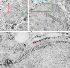
Vinyl chloride and low Vit C exposure: guinea pig, liver, mitochondrion, intramembrane, inclusions — a long title. This is an electron micrograph from the liver of guinea pig # 7 in a study about inhalation of vinyl chloride in conjunction with / without vitamin C. Dr. Martha Radike (who was a wonderful friend and colleague) did this study back in the early 1980s, but I don’t believe she ever published the results. This particular animal (M8029) inhaled 600 ppm vinyl chloride 5 days a week for a year. The specifics of this which I cant remember (30 years ago) are 600 mmp VC, 4 hours a day, 5 days a week for a year, and this particular animal received 2-10 mg of vitamin C per day. The mitochondria in lung, btw, were also very enlarged, though at this point I did not see any of these little inclusions within the mitochondrial membrane.
 Published reports have described changes from vinyl chloride exposure in liver: hypertrophy and hyperplasia of hepatocytes, activation and hyperplasia of sinusoidal lining cells, fibrosis of the portal tracts and the septa and intralobular perisinusoidal regions, sinusoidal dilation, and focal areas of hepatocellular degeneration. While the latter came out of a document that said DO NOT REFERENCE DO NOT QUOTE (oh well, excuse me), these are separate observations, but supportive of the unusual changes that Martha Radike (and I) found as mitochondrial changes seen in liver her year-long vinyl chloride exposure experiments in guinea pigs. There is a single mitochondrion pictured here, and two enlargements of areas (red boxes designate insets) which show the most wonderful inclusions I’ve seen twixt the outer mitochondrial membrane and inner mitochondrial membrane (I guess I could look for it in the intra-cristal membrane too but I haven’t yet). There are some misshapen mitochondria with cristae showing such in the air-breathing controls, which I will post, that received higher doses of vitamin C. One wonders if the health of the entire colony played more into the results of all these changes (lung and liver both).
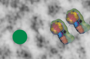
These little lollypops stuck on either side of the membrane are neatly alternating in a wonderful order and speak to something that I have not investigated but which was immediately apparent when I did googled “lollipops and inner mitochondria membrane” this may be ATP synthase. The dimensions are close to a reference I found: Structure of the mitochondrial ATP synthase by electron cryomicroscopy by John L. Rubinstein, John E. Walker, and Richard Henderson. There are some great videos on this molecular machine: HERE and HERE and HERE In the bottom section of the top micrographs the RED dot is @27 nm (ribosome), green dot @ 18 nm, bar marker, 100 nm. In the small bottom image i just cut and pasted two visuals of ATP synthase onto an enlarged portion of the above micrograph. I added two charts to the pdf uploaded previously describing the materials and methods for vinyl chloride exposure. Find that document –> vinyl_chloride_vit_C_guinea_pig_lung1.
Next post is going to be a few pix of the ribosome bundle which is adjacent to some of these portions of intramitochondrial membrane with ATP synthase strings.