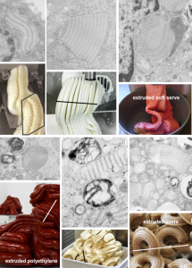
The granule in the alveolar type II cell of guinea pig, ferret and dog is unique, in that it appears to have wonderful shapes, concentric, U-shaped, linear, branching (even highly branching mostly at perpendicular planes to the original layering). I was trying to figure out what could cause such variation in shape. One keeps in mind that these are cross-sectional images, 2D of the 3D cytoplasmic granules, and therefore can be difficult to interpret as a whole granule.
To note first: there are often parallel “jets” (which I have called periods, just to be proper ) for the production of surfactant protein A (yep, I am calling it that with nothing but circumstantial evidence). Sometimes there are 10 or 15 such nidi of production, each perpendicular to the long axis of the layering (banding) and having about 4 ribosomes (the 4th ribosome in each period serves as the 1st in the adjacent layer). The amount of resistance at the opposite end of the granule to protein synthesis, and the rate of protein synthesis (at the growing end(s) of the granule where the ribosomes are ) will determine whether this “jet” will spew out SP-A in an unhindered stream to create a linear granule or whether it will buckle, twist, spin or curl.
I began searching extrusion images on google, and these below (easily-found set of 6 extrusion images which illustrate the point) do a really good job of matching what is seen electron microscopically, of what becomes of the structure of protein layering during SP-A production when the protein produced meets with resistance. Awesome… ha ha, a natural physical phenomenon, I will bet. What scientific journal will publish my diagram… ha ha, probably not. Too funny.
