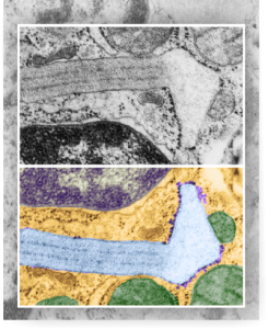
…and yet another cover submission for the manuscript on SP-A granule and electron microscopy. This one is a little more dramatic than yesterdays post from the bottom right part of the image. I like it better. It shows the quintessential granule, in black and white at the top, color at the bottom in a vertically mirrored image, directional ribosome attachment, electron-lucent areas under the non-oligomerized protein part, distinct periodicity to the dense bands (three complete “periods” in the lengthwise portion of this granule) and also the periodicity to the dense layers (3-4 dots/100nm) and even some of the faint periodicity found in the central lighter layers. In this electron micrograph one also sees part of the nucleus (bottom), a couple of nuclear pores, and some perinuclear space (but no granule formation in the perinuclear space is seen in this image, but it is found there frequently) and on the top, portions of three mitochondria and one adjacent to the lucent area of the granule. There are also other profiles of RER, coming and going which at some point might connect up to layered putative SP-A protein granule structures.
