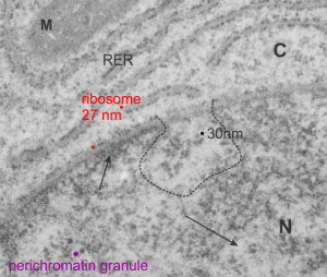
From a study in the 1980s looking at exposure of pregnant dams to CO or dichloromethane by inhalation, here is a kind of “pristine” hepatocyte from ad gd 20 fetal rat. The nuclear pores are numerous but not really that distinct, and the RER is nice and flat but also has a dense content, and the mitochondria have cristi that are not completely flattened and there is not a lot of it, and very little SER that I can find and this cell is adjacent to two hematopoietic cells, no desmosomes between. M=mitochondrion, C=cytoploasmic side, N=nucleoplasmic side arrows point to condensed (closed) and euchromatin (open). Ribosome can generate a relative size comparison for other structures, being about 27 nm in diameter (red dots), the nuclear fibrils just at the nuclear pore (almost dead center in the micrograph and within the black dotted lines) about 30 nm (black) and perichromatin granule in purple. The project, as I recall, did not uncover any differences between the exposed and non exposed animals, and in this case, the hepatocyte looks nice.