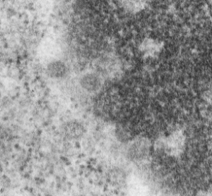
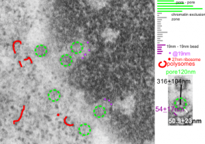
Three measurements taken in a series. Pore to pore, pore to to edge of chromatin (which i found is called the chromatin exclusion zone, so am sticking with that since it clearly is an active event, and may vary with cell type and cell activity) and inter-@19 nm “beads” that are seen as the organized chromatin just adjacent to the nuclear pore (the first thing seen after the chromatin exclusion zone). This micrograph is from the same images as a previous post, which is 16027_65718_14Cos_ko 24hr no NTBC measurements.-2a.
 n=5, pore to pore=316nm+104nm; n=15, chromatin exclusion zone, 50.9+23nm; n=5, 19nm to 10nm chromatin granule, 54nm+17.7nm