Category Archives: Ultimate order, the cell

The beauty and order of life is astounding.

Kudos to Flying Pig Marathon recycle – repurpose – reuse efforts

THIS IS THE KIND OF INFO ThAT WE ALL NEED TO SEE DAILY
Reduce, Reuse and Running

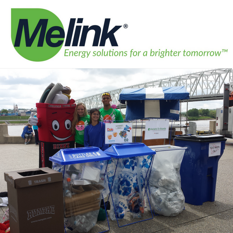
Thanks to the incredible help of all participants, the Green Team volunteers, and staff, the 2017 Flying Pig Marathon weekend of events diverted a total of 78% of waste, exceeding our 2017 goal of 75%. We were able to successfully reduce our carbon footprint in numerous ways, including recycling heatsheets into Trex decking and railing products, recycling food wrappers through TerraCycle, recycling unused medals and participate bibs, and repurposing medal ribbons as lanyards or recycled through Goodwill’s textile recycling program. Banners, discarded clothing at the start, and leftover food were all donated to local charities. By offering carpool parking, we reduced the number of cars on the road by 1,194. We also offset part of our carbon footprint by partnering with Taking Root in planting 5 trees along the Pig marathon course.

The Flying Pig Marathon is excited to announce a partnership with Melink Corporation of Milford as the “Official Sustainability Sponsor” of the 2018 marathon weekend.

“We are thrilled to have Melink join the Flying Pig in our sustainability efforts,” said Iris Simpson Bush, Executive Director. “The Pig and its year-long events have made sustainability a prime focus for a decade now, and Melink will only help continue our efforts to preserve the environment.”

Intermediate filaments: the great connectors between and within

I dont know exactly how to go about diagramming this…. but it seems that the filaments (no surprise should be had) are so critical….  for so much in cell function, just in orchestrating all the comings and goings of the organelles, in addition to some filaments making sure what enters the nucleus through those cute little nuclear pores and regulating the presence of mitochondria (energy, calcium, apoptotic proteins and membrane potential) at nuclear pores and at desmosomes (maybe adherens junctions as well).  All in all, a highly complicated and interesting topic.

here are a couple of better diagrams i found on google


Desmosomal – mitochondrial tethers: examples in ciliary bodies

148nm from plasmalemma to mitochondrial outer membrane, they measure 63 to 84 from the outer limits of the desmosome (a hard place to judge), and they note that there is a lucent area (of about 5 nm, or half the diameter of an intermediate filament) between each of the dense lines of the intermediate filaments and the mitochodnrion.
41nm is intercellular space of the desmosome.
815nm=length of this particular desmosome (approximate).
Annulus=about 74nm of plasma membrane increased density on either side of the desmosome.
3 to 5 intermediate filaments parallel to mitochondrion and desmosome.
Schlötzer-Schrehardt et al 1990 also mention that there can be just one mitochondrion with one desmosome on one cell of the two involved, or there can be bilateral symmetry (i.e. one mitochondrion on one desmosome in each of the attached cells, or strings of desmosomes linked by one single mitochondrion…or a mirror. They show an image (below) of eight tethers, two mitochondria, five desmosomes, the latter spaced variably (they measured 50 – 350nm). They also remarked that when there were multiple tethers, that the intermediate filaments were more prominently arranged.

28nm=intercellular space of these desmosomes
113nm=mean length (n=5) of this particular desmosomal set, longest is about 168nm (that would be something less than the anticipated measurement of the diameter).
70nm from plasmalemma to mitochondrial outer membrane (mean of four measurements).
63nm=annulus
4 intermediate filaments parallel to desmosome and mitochondria in one opportune space (lower left of this micrograph).
Schlötzer-Schrehardt et al 1990 commented of the possibility of calcium being one of the reasons for such tethers.  Aside from the anatomical location such a large frequency of desmites providing clues as to the energy required for rapid and frequent adjustment of the iris, and the likely stress on the cells…. the need for an energy resource is pretty obvious, as would be the need for releasing stretched areas from their bonds.

I wonder if ciliary processes represent a tissue with extraordinary desmite adaptability and pliability. I will work to produce an excel file with all the tissues I can find, and notations (or actual counts as provided in the Schlötzer-Schrehardt et al 1990 publication.


Early article on desmosomal-mitochondrial complexes

After so many years of looking for articles on this topic (I actually wondered how this phenomenon could go unnoticed for so many decades) i did find one manuscript (which references two others) by Deane et al 1966 which mentions them and shows several micrographs (sometimes pretty low manification). They found these first in fetal and neonatal tissues, so this is a clue and also important for the concept that I have which is that in tissues with higher rates of cell division that also are epithelia which connect at environmental interfaces (meaning the “outside” whether that is physically outside or not) have an active mechanism for building and disassembling desmosomes.  They felt it was a one-way passage…. i think it is more a modeling-remodeling phenomenon.  So this was nice but bitter sweet at the same time since I thought my early paper in a very obscure journal in the 1980s was the first.

(I am going to use this acronym…desmosomal mitochondrial tethers  regardless of how silly it sounds just because it is too time consuming to type out 32 characters when 7 will do, my apologies.)

miller et al, desmosomal mitochodnrial interactions_1985
deane_1966_desmosomal mitochondrial complexes
In figure 1, and enlargement inset called figure 2 (a portion of which is below) of their article, they make a cute comment…. about a desmosome beginning to develop nearby to a reasonably well defined desmite. It would be better perhaps to have recognized that there were two possibilities — one, that it was a tangential cut to a sequentially placed tether along a single mitochondrial outer membrane, and the other that it was a desmite “being built or broken”. This little area of not completely well defined junction they label with a black arrow. You can see that it is not quite a desmosome and the mitochondrion really isn’t that close to it.  The label TF (i don’t know why it is in italics) surely stands for tonofilaments…. which I refer to as intermediate filaments.


Just using their image and their micron marker (0.5mu = 500nm) it seems that their cut in this desmosome is just off center but close (diameter with the annulus) at about 286nm (central cut yields a desmosome of 300nm) and a diameter of 214nm excluding the annulus.  About 162nm span the distance from the outer mitochondrial membrane to the plasmalemma membrane of the desmosome of the cell on the same side. A quick measurement for the thickness of the extracellular space of the desmosome in this image was about 18nm.  About 15nm /2 for the dimension of the annulus area on either side of the desmosomal spot. This micrograph shows unequal annulus dimensions.  Lovingly I point out the scratch (likely on the old acetate negative) lower left side of the desmosome… and in sympathy say, my negatives and micrographs have many such scratches. One thing to not is that 3-5 intermediate filaments are lying almost parallel to the plasmalemma and the outer mitochondrial membrane… certainly NOT like the diagrams seen routinely  — see my post with desmosomal diagrams with hairpin (wrongly directed) lines for intermediate filament attachments to desmoplakin molecules.

What we don’t know we either fear or worship!

What we don’t know we either hate or worship! How strange this is, yet for eons this has been true. I was making a crossword puzzle, using the concept of heaven and how oddly this place-space-destination-resting spot-culmination-equilizer-paradise concept has changed over time. I ran onto the concept of JNN or jinn or jinni or anglicized as genie, and found this gorgeous page from a manuscript.  The jinn were important god-like beings at one point near pre-history but were demoted to human like then smaller and smaller unknown forces for events and ailments that were not understood.  All dentists will totally love this image of two teeth, with the pulp and root canals occupied by jinn….  causing a tooth ache.

As soon as the elements causing teeth to rot and die and ache, jinn went on to become some other force, no longer hated nor worshiped.  We are a strange species at best. Thank you wikipedia.


Desmosomal symmetry: not a complete mirror images side to side

Desmosomal symmetry: not a complete mirror images side to side, since it seems to occur in my micrographs, and obviously too in this micrograph from Green and Gaudry, that the separate cells (adjacent cells bound by the single desmosome) may have a propensity to have intermediate filaments coursing by at perpendicular angles.  Maybe also chance – I guess it is a 50 50 chance to be chance (haha).

But also in this quote from Green and Gaudry.…I take exception to their remark that the desmosome is tripartite. A quote from their paper yielded this text of which several parts make no sense to me “electron micrograph further illustrates the highly organized ultrastructure of a desmosome (yes, i agree) in which mirror-image (partly agree, but not always true), tripartite electron-dense plaques (show me 3, ha ha, and are you counting lucent bands, plasmalemma (not really to be counted and intermediate filaments?) sandwich a central core consisting of adjacent plasma membranes (wait, plasmalemmas are NOT part of the central CORE, and what is meant by CORE anyway, that is a term that needs definition) bisected by an intercellular zipper-like midline (yes, zipper like dense midline -i agree)

They didn’t take into acount the different directions (which may be purposeful) of the intermediate filaments coursing by the desmoplakin molecules in the adjacent cells… as sometimes they appear as 10 nm cross sections and other times as low arcing swooshes. I ask, what part of the plaques are divided into 3??? This is a confusing, since there is nothing distinct about the 3 parts they refer to… they could represent many/or any different layers of this organized structure. Their own electron micrograph (pasted below) shows pretty convincingly that there is not total mirror symmetry to the desmosome since their cell on the bottom part of the image shows cross sections of intermediate filaments while the cell on the top part of their image shows the longitudinal swoosh of intermediate filaments.

One thing that their electron micrograph shows that is rarely commented on is the desmosome annulus… this ring which of plasmalemma which is just slightly morphologically different than plasmalemma further from the desmosome.

While on the topic of their diagram, i think more care could have been extended to the depiction of the tight junction which is really a weld, and they show it as a wider structure than the two blended plasmalemmal membranes really are. And the adherens junctions are diagrammed to be as prominent as desmosomes, which they are not, and the labeling of adherens junctions actually appears closer to their green blob desmosome and thus doesn’t really direct attention to the junction they are wanting to show.  Maybe that works for a visual aid for some, but for me it does the opposite…. working from a place of knowing the actual structures and trying to figure out what visual errors the “diagrammer” has created… is frustrating.  This brings up the legion of diagrams of really poorly drawn desmosomal structures… ha ha.  I do wish more care could be taken in scientific illustrations..N
Not a single diagram that I have seen on desmosomes and intermediate filaments has mentioned the importance of the presence of mitochondria in building and breaking down these structures.

Different intercellular joints?

Weld (stronger than rivet)*; light in weight; can be made with access to one side only; making and breaking requires little energy= tight junction
Rivet (loss in strength); some flexibility; heavier; requires access to both sides (2 cells); making and breaking requires energy; = desmosome
*http://www.1920-30.com/architecture/rivets-welding.html : this source says there is no loss of strength with a weld, but particularly, when i viewed micrographs of spot welds, sometimes the sheets of metal look thinner and the texture of the metal has changed under the electrically produced weld.

The desmosome is not really a butt joint, since the center plate in a butt joint is the intercellular space and there is no edge to be joined in the respective adjacent cells. Maybe it could be considered more like a double-rivet lap joint. Or a double-rivet double strap joint.

Environmental impact on desmosomal joints are: tack ( how easily a bond is made and broken  ); peel (the force needed to break the bond between two cells – including peel angle and direction); shear (sliding between surfaces), and then there is the list of molecular forces (Ions, enzymes, water, heat and energy).

Here is an interesting quote from label makers, “Non-carboxylated emulsions tend not to build adhesion over time, an important attribute in removables. Tg influences pliability, especially at low temperatures. (Think labels applied in the freezer section). Gel content is a measure of cross-linking within a styrene-butadiene emulsion polymer. Less cross-linking results in higher tack, but more cross-linking provides higher shear. If necessary, a tackifier can be added to the emulsion to raise tack even more.” and one wonders how this polymer science stuff relates to the polymers in desmosomes.

The desmosome, the adherens junctions — both have attributes, there is a  trade-off from one type to the other, for different jobs and  modifications  within each category.

Shear testing for desmosomes, as layered structures,  disruption along the plane of the cell membranes. I guess a desmosome can be called a viscous adhesive.  haha. or a velcro spot.

Desmosomes: ductile, double-sided, shear load rivets

Desmosomes are kind of like Oscar rivets, and are blind perhaps, or double-sided. the latter have splits (the desmogleins and desmocollins) along the shafts. These splits cause act like flares and join in the central dense line (periodicitities) of the desmosome. The plakoglobins and plakophilins form the “head” of the rivet along with desmoplakin, and these rivets are unlike most rivets found for metal and wood work, because they are flexible, and have a  “spring-hinge” at their head, the desmoplakin-intermediate filaments junction (which i believe is perpendicular not parallel (as most of the diagrams show), which can flex and bend and allow for motion of the desmosome as it gathers together as weld between two cells.

Below is a set of diagrams taken freely from the internet from publications on desmosomes (including my own). It can be seen that the majority of diagrams don’t really “see” what the microscopist sees as the course that intermediate filaments take when they are adjacent to desmoplakin.  The diagrams on the lower left, and all those in the center and the lower right have intermediate filaments coming from the cytoplasm and making hairpin turns at the desmoplakin molecules…. However, the micrograph on the upper left, and the diagram in the upper right have the intermediate filaments which actually appear to arc ever so slightly, to be NOT forming hairpin turns to desmoplakin, but gently curving perpendicularly to the desmoplakins.

It seems more likely that the type of shear stress (the connection between desmoplakin and the intermediate filaments perpendicular to one another) would be more advantageous at binding two cells together than the diagrams below (with hairpin turns) which suggest that the connections between intermediate filaments and desmoplakin are parallel, thus involved in tension stress.  The former intuitively suggests more efficient shock absorption over a smaller distance than the latter, and actually looks more like the actual anatomy than the latter.

The disparities among the images from electron microscopy and the diagrams from the literature are the reason that I began to try to think this through. Intermediate filaments are seen in micrographs that look perpendicular to desmoplakin (like what is seen in the micrograph in the upper left part of the diagram as brown lines), or as 10nm round dots (if they are cross sectioned).  Any ‘apparent’ long intermediate filaments parallel to desmoplakin (as seen in the bottom center electron micrograph) can almost always be explained as tangential cuts.

This arrangement is most clearly demonstrated when intermediate filaments form a flat perpendicular band between the desmoplakin molecules of the desmosome and the mitochondrion when the desmosome and the mitochondrion are tethered together.
diagrams of desmosomes

Desmosome dimensions relative to adjacent membranes and mitochondria

It seems to me that there is a pattern of thickness  changes(width or height if you wish… because of the orientation of the diagrams below) and the rigidity of the plasmalemma inherent in the desmosome (likely due to the transmembrane parts of desmoglein and desmocollin ) and that rigidity includes a specific intercellular space dimension. I have seen this published at about 34-38 nm (that is, from the inner leaflet of the plasmalemma of one cell to the inner leaflet of the plasmalemma of the adjacent cell) to be something on the order of 38 nm.  Using that dimension, if i measure from inside plasmalemma of one cell to the adjacent cell and compare that to the width from the same places in the 200 nm ring or annulus around the desmosome there is going to be a change in the dimensions (which i could measure as just a few nm greater than that within the area of the desmosome proper. And, then a reduction in the intercellular width (per the more routine proximities of two cells) which becomes something like 40 nm.

Here is an article which suggests some dimensions for the desmosome, but does not address adjacent variations in the plasmalemmae including the anulus.  You can compare with two images and measurements below. Keep in mind that the two images below are not derived completely randomly, as I picked two which had substructure which was clear enough to measure.  The annulus of these two desmosomes is indistinct, and longer than that seen in other desmosomes. This likely relates to the plane of section on a perpendicular axis of a round desmosome and surrounding annulus (the latter being increasingly seen as one sections nearer the periphery of the actual desmosome).

top diagram shows a length of two adjacent plasmalemma, measurements relative to that image, bottom one is a different desmosome, notice brown lines for intermediate filaments, black line for length of the desmosomal spot, dotted lines at periodicities of the bridges between desmocollins and desmogleins, outer dense line with dots, periodicities of the extracellular membrane anchor, pink bracket, thickness of the desmosomal intracellular elements, which includes proteins of the intracellular anchor (desmocollins and desmogleins) plakoglobin, plakophilin, desmoplakin and intermediate filaments (visible at the desmosomal-mitochondrial tether on the bottom more clearly than the desmosomal-mitochondrial tether seen at the top of the lower micrograph.  While a big deal is made of the inner and outer dense plaques of the intracellular part of the desmosome, the lower portion of the lower micrograph doesn’t make that case.  Were the micrograph sectioned end on to the intermediate filaments below, there might be a more visible inner dense plaque.  The outer dense plaque (plakophilins, plakoglobin portion and desmoplakin proteins) is well defined. NB, there is a “flatness” or “rigidity to the outer mitochondrial membrane where the intermediate filaments lie beside it… i hope to search for proteins that might be involved in the linking of mitochondria and intermediate filaments.