Category Archives: Mitochondria: electron microscopy

odd mitochondira, great mitochondria, electron microscopy and ultrastructural details

Where is the list?

Just experiencing info-disorganization-overload… ha ha

Trying to do a simple thing… to list and learn about the proteins in mitochondria.
This should be a simple task. I am looking for all proteins known to abide in the inner and outer mitochondrial membranes and those segregated to the cristae junction and the cristae themselves.

Everything…. Everything has an acronym, inconsistent and often just silly, nothing is synthesized into a neat package or publication.
I can accept that not all proteins are known at this time, but it is harder to accept that random bits of important information are not synthesized neatly into a package…
by kingdom
by phylum
by class
by order
by family
by genus
by species…
by tissue…
by cell type
by membrane position
by function
fun graphic a mitochondrionpseudocolor mitochondrion

More on Form follows function in mitochondrial cristae organization

Not much text with this post, but here is a highly simplified, not real detailed, summary of what some think about mitochondrial cristae morphology. This is very basic, i intend to make some really nice diagrams including the inner mitochondrial membrane proteins as represented in 3D by the protein databases. That will include the OSPHOS proteins, and the channel and transport proteins and the cristae junction proteins.  But for now…. and blatantly missing several types of cristae (triangular in particular) here is a line diagram of cristae and some associated properties. The electron micrographs on the “aerobic” side of the diagram are from various images of hepatocyte mitochondria just cut out of existing micrographs, on the anaerobic side, the mitochondria are selected from isolated mitochondrial fractions. Both are traditionally prepared TEM processes. Abbrev: IMS, inter-membrane space; white rim area. Outer and inner mitochondrial membranes=black lines. Grey interior= mitochondrial matrix. Micrographs, not all the same scale,


While i love the tomographic 3D reconstructions of portions of the cristae, it seems that this is sort ofwhat most of us rendered in our minds before those data confirmed the structures.


Mitochondrial cristae junctions and pores?

Searching through micrographs to determine whether or not i can visualize a mitochondrial pore or crista junction.  Here is a cutout of a border of a mitochondrion which looked to have several adjacent and more or less evenly spaced cristae along one outer mitochondrial membrane. None of these exposes a clear connection where an inner mitochondrial membrane space should appear. Also of note (in this experimental set of animals) there tends to be a narrowing at the base of the cristae, and a little ballooning of the remainder of the cristae bring. This might be a manifestation of the cristae junction. In a stretch of outer mitochondrial membrane about 475nm in distance there were 6 areas where the former and the cristae membranes meet.

In this particular mouse liver electron micrograph the animal was a hepatocyte specific ko for GCLC, specifically negative 18408 block 78375 wc/ii animal#5 and 50 days old. red dot=@27nm (one cytoplasmic ribosome) used as a micrometer.

mitochondria liver electron micrograph cristae junction
Looking at the matrix of this mitochondrion several densities appear which may be too small of mitoribosomes.


Another mitochondrion which i examined and pseudocolored the same those above, has about 1 cristae membrane junction pore per 250nm or so counted over a stretch about five times that long. Clearly, physical state (these KO mice) nutrition, gender, cell type and function which the mitochondrion is supporting.  Green is the inter cristae membrane space,  pink is the outer and inner mitochondrial membranes, blu background is the mitochondrial matrix. The tissue from which these sections were made was a ko, (conditional KO of GCLC in liver).

Isolated mitochondrion

Just looking for the myriad features that should show up in mitochondrial ultrastructure. THis isolated mitochondrion is from a wild type mouse post natal day 14, liver.  It does show areas where cristae membranes (inner mitochondrial membrane) and outer mitochondrial membranes create the inner mitochondrial membrane space (green) and the membranes themselves (pink) and the matrix (blue).  Within the texture of the matrix it is tempting to try to find circular mDNA and mitoribosomes and there are hints of them here but nothing really obvious shows up. Trying to find some difference in inner mitochondrial membrane density which would indicate the crista junction is pretty disappointing as well.  Not all structures named, or presumed to be real, can be found in routine micrographs. There is one little circular structure in the middle of the matrix which I might well have included as cristae membrane. isolated mitochondrion cristae inner membrane

Crista junctions in mitochondria

I am trying to visualize crista junctions in mitochondria in the liver (mammalian). These might be visible here, in a KO mouse which has increased oxidative stress (previous posts). It looks to me like there is a small consistent area at the place around cristae approaching the outer mitochondrial membrane that looks “different”?  Find the 100nm markers (vertical at the edge of a crista and horizontal by the red dot over on the right hand side) are an approximate marker for the size of a cytoplasmic ribosome (27nm) and calculation for 100nm from that.

Cristae in this particular mitochondrion and many others show an increase in the amount of matrix space and a more vesicular type of cristae, and some times have cristae inclusions.

Mitochondrial ultrastructure

It is a little difficult to get the whole picture of mitochondrial ultrastructure together in one place and in one image. The the large and complex groups of proteins like ATP synthase,  or mDNA and mitoribosomes and the proteins for energy conversion, or calcium storage, ion transport, proteins that are involved in cell growth, division and apoptosis, as well as the basic mitochondrial shape and volume density of and shape of cristae and the amounts of mitochondrial matrix, and many other things of which I am sure i am not aware….are difficult to sort out.  Ultrastructural aspects related to these functions are not well understood, and at best poorly diagrammed.  I have found one website (biology by the numbers) which does do a great job of labeling size and giving measurements of some aspects of mitochondria, but of course not all, and not neatly organized. There really probably is not going to be much consensus about these variations in mitochondrial ultrastructure because the influence of tissue, cell, metabolic state, types of fixation, the embedment, as well as many variations in scope mag, deliberate manipulation of the images, poorly kept records, presumptions, and the list is endless.  So the question is how does one go about diagramming (or imaging) the most educational presentation about the mitochondrion. A really perplexing structure is the “pore”  or cristae junctions.

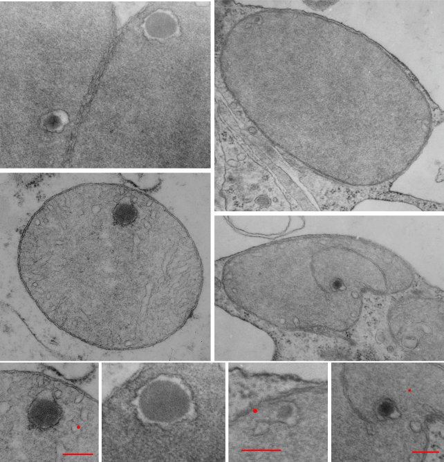
Add to that list of  variables, the long list of diseases where mitochondrial shape is found and the concept of imaging the “perfect educational” mitochondrion becomes more difficult.  So trying to find a great diagram came about because of an inclusion that I found in the cristae of some of my own micrographs.

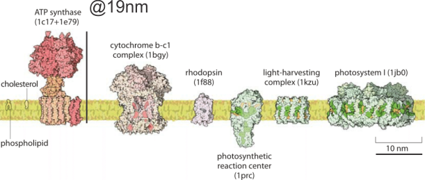
It would just be nice to see these structures ( 5-10nm diameter) in the literature with the normal ultrastructure of the mitochondrion, which is not really that well documented for standard TEM images though there are some nice molecular diagrams of membrane proteins.

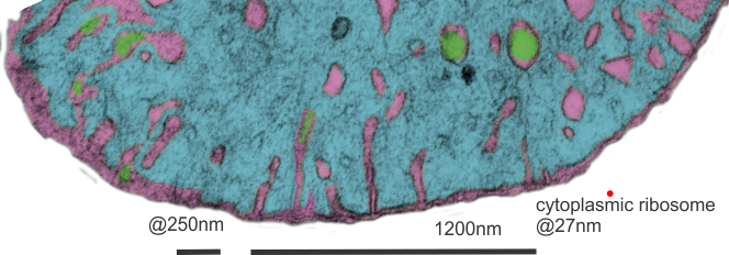
The round electron dense inclusions are unknown (in a crista space)(in these not-perfect micrographs). It is possible that in the bends of a couple of cristae, there are densities right at the bends which would seem to be identifiable.  Looking at the blue dots in the top image below, right at the curvature of a crista–one might see those dots as ATP synthase (blue dots are given at the approximate size that ATP synthase should be ( “biologybythenumbers” diagram (thank you for that) (bottom image).  Also, separate, larger orange dots could be mitoribosomes which reportedly are just smaller than cytoplasmic ribosomes (picture as red dots (taken from the same micrograph as seen on the left). A second reason for working on mitochondrial ultrastructure is to try to figure out what the electron dense (and homogeneous round) protein is within this crista space)

Metal? cristae inclusions in mitochondria

I found a paper that showed a TEM of a dense round intracrista inclusion that had some similarities to what is seen in the Gclcwc/ii hepatocyte specific KO mouse at 50 post natal days.  Paper is Characterization of Intracellular Inclusions in the Urothelium of Mice Exposed to Inorganic Arsenic Toxicological Sciences 137(1), 2013 by Puttappa Dodmane et al. I cut and pasted part of one of their electron micrographs of a mitochondrion with such an intracristae inclusion (right side of picture) next to what is seen in the KO mouse (my picture, left side of image).


What kind of diagram is this? mitochondrial cristae

I find that diagrams and illustrations for science are so often NOT GOOD, NOT EXPLANATORY, and are TOTALLY CONFUSING.  Here is an example. It is from a very nice article, (Biochimica et Biophysica Acta 1793 (2009) 5–19), but the diagrams of the arrangement of cristae just doesn’t make visual sense.  It is an injustice to readers to create confusing images and put them up as learning aids.  THis diagram misuses the shading when it creates a look of 3dimsnsions…. it makes no visual sense in any of the three drawings in this figure.  To create shading on one portion of a diagram (e.g. the middle figure) which in this case was the intercristae space, which is called the intermembrane space, and NOT make the shading equivalent on the mitochondrial matrix is just careless, and misleading. The lower diagram is beyond deciphering…. it looks like there are three orange fingers poking up, from a space with holes.  Nothing resembling anything that is shown with real TEM images.  So sad.  It wastes time and  sends wrong information.

Mitochondrial intracristae-space inclusions

Mitochondria provide the cell with energy as well as integration of metabolic pathways for biosyntheses of heme, iron–sulfur clusters, and nucleotides, triggers for cell apoptosis, and reactive oxidative species signaling and includes a circular mitochondrial DNA (a few thousand copies) of about 16.5kb which encodes for some peptides which are required for oxidative phosphorylation, which are transcribed and translated to mt-mRNA, mt-rRNAs and 22 mt-tRNAs, and their own mitoribosomes within the mitochondrial matrix. Many other protein (several hundred) are encoded by nuclear DNA, transcribed, modified in the cytoplasm, then imported into mitochondria. Nucleoids (the mtDNA and associated proteins are ascribed a size (not by me) of something around 100nm) (which means they should be visible in routine transmission electron microscopy.
Looking through many micrographs from my own collection i found several which had unique inclusions within a membrane space (maybe a crista space) which reminded me of the fine texture of DNA in an apoptotic cell which had been digested down into the 2000 to 250bp fragments (laddering of nuclear DNA), actually maybe even finer texture. red dots=approximate size of a cytoplasmic ribosome, bars=approximately 270nm. several micrographs derived from 2 experimental mice, hepatocyte specific knock out of the Gclc gene. 50days old. Mitochondrial changes have been reported (HEPATOLOGY 2007;45:1118-1128) in these animals previously, but intra-cristae inclusions were not reported. These mitochondria have an extreme expansion of the matrix area, pressing cristae to the sides, of the mitochondrion.  Interestingly, there really isn’t a lot of substructure to the matrix (as one might expect some mitoribosomes or mtDNA clusters