Category Archives: Mitochondria: electron microscopy

odd mitochondira, great mitochondria, electron microscopy and ultrastructural details

Desmosome dimensions relative to adjacent membranes and mitochondria

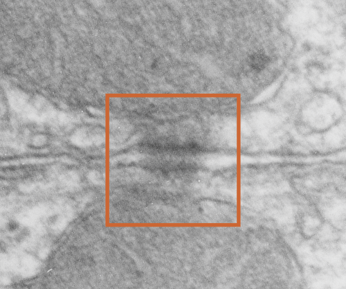
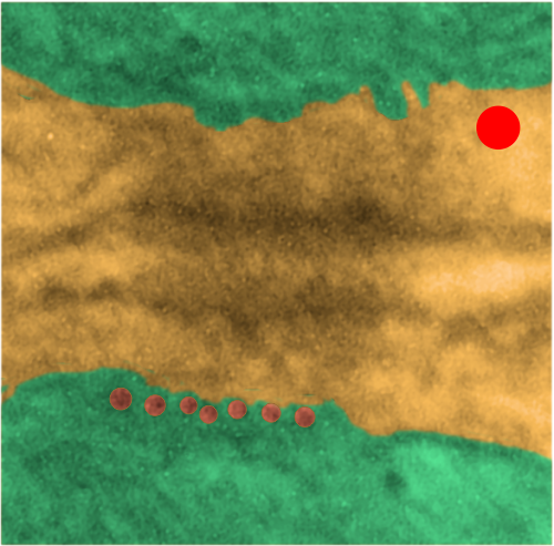
It seems to me that there is a pattern of thickness  changes(width or height if you wish… because of the orientation of the diagrams below) and the rigidity of the plasmalemma inherent in the desmosome (likely due to the transmembrane parts of desmoglein and desmocollin ) and that rigidity includes a specific intercellular space dimension. I have seen this published at about 34-38 nm (that is, from the inner leaflet of the plasmalemma of one cell to the inner leaflet of the plasmalemma of the adjacent cell) to be something on the order of 38 nm.  Using that dimension, if i measure from inside plasmalemma of one cell to the adjacent cell and compare that to the width from the same places in the 200 nm ring or annulus around the desmosome there is going to be a change in the dimensions (which i could measure as just a few nm greater than that within the area of the desmosome proper. And, then a reduction in the intercellular width (per the more routine proximities of two cells) which becomes something like 40 nm.

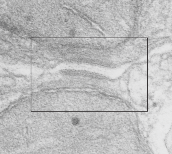
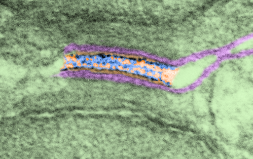
Here is an article which suggests some dimensions for the desmosome, but does not address adjacent variations in the plasmalemmae including the anulus.  You can compare with two images and measurements below. Keep in mind that the two images below are not derived completely randomly, as I picked two which had substructure which was clear enough to measure.  The annulus of these two desmosomes is indistinct, and longer than that seen in other desmosomes. This likely relates to the plane of section on a perpendicular axis of a round desmosome and surrounding annulus (the latter being increasingly seen as one sections nearer the periphery of the actual desmosome).

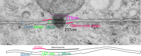
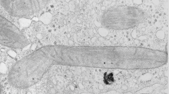
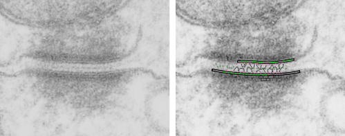
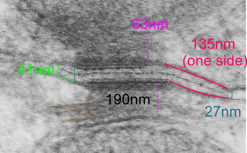
top diagram shows a length of two adjacent plasmalemma, measurements relative to that image, bottom one is a different desmosome, notice brown lines for intermediate filaments, black line for length of the desmosomal spot, dotted lines at periodicities of the bridges between desmocollins and desmogleins, outer dense line with dots, periodicities of the extracellular membrane anchor, pink bracket, thickness of the desmosomal intracellular elements, which includes proteins of the intracellular anchor (desmocollins and desmogleins) plakoglobin, plakophilin, desmoplakin and intermediate filaments (visible at the desmosomal-mitochondrial tether on the bottom more clearly than the desmosomal-mitochondrial tether seen at the top of the lower micrograph.  While a big deal is made of the inner and outer dense plaques of the intracellular part of the desmosome, the lower portion of the lower micrograph doesn’t make that case.  Were the micrograph sectioned end on to the intermediate filaments below, there might be a more visible inner dense plaque.  The outer dense plaque (plakophilins, plakoglobin portion and desmoplakin proteins) is well defined. NB, there is a “flatness” or “rigidity to the outer mitochondrial membrane where the intermediate filaments lie beside it… i hope to search for proteins that might be involved in the linking of mitochondria and intermediate filaments.


Mitochondrial-intermediate filament functions… only one at a time? probably not

I found a nice review article the title is “Intermediate Filaments as Organizers of Cellular Space: How They Affect Mitochondrial Structure and Function by Nicole Schwarz * and Rudolf E. Leube”  free online, about intermediate filaments and mitochondria, which is part of my reference list as I am trying to figure out where there are species differences in the ultrastructure of desmosomal-intermediate filament-mitochondrial tethers.  So this is a nice diagram but i think it is too primitive, or perhaps too limited, or maybe just has not been taken to the next step.

This image is their Figure 1 which shows three possible intermediate filament – mitochondrial arrangements.  My exception to it is the following : there are areas around each mitochondria where all these types of bonding-binding-exchange take place, at least it seems to me.  For instance, the binding of intermediate filaments to the outer mitochondrial membrane (OMM) probably doesn’t occur (at least in hepatocytes) in a complete manner surrounding the entirety of the outer mitochondria membrane, but rather definitely is “focal” as it looks when it is tethered to another cytoplasmic organelle such as the outer edges of the nuclear pore, or the inner plaque of the desmosome.  There is a definite change in the structure of the outer mitochondrial membrane, gross enough that it is seen with TEM, and looks like a thickening of the OMM and a “flattening” out of that area which is engaged with intermediate filaments. There is also a shape change to the mitochondria when they are bound (i prefer the word tethered) to the filaments.  (so still talking hepatocytes here). Intermediate filaments positioned remotely from the OMM and called “signaling” is something that I don’t believe would be seen physically.  As for confinement, there are certainly areas of confinement of organelles, and they might be called “no fly” zones, I also need to find an example where the mitochondria are almost repelled by intermediate filaments.

In a couple of mouse models i have looked at, intermediate filaments themselves can be repelled (or it looks that way), of which one is the central confinement of intermediate filaments in small intestine microvilli  where the gastric HKatpase is missing).  There are a dozen, at least, intermediate filament x OMM sites that can be seen hepatocytes where there is obvious “tethering”, and i bet signaling as well.  So these categories might have a lot of crossover.

I am thinking that in order to signal, the intermediate filaments need to be physically closer to the mitochondria than this diagram projects…. I wonder how that could be determined with TEM.


Questions about desmosomes and mitochondrial junctions

Desmosomes:

(who names these proteins — desmosome..  desmo — and soma, that is the easy button, cadherins…  well calcium dependent adhering molecules, i guess thats ok, desmoglobins… not so good, plakophilin, maybe that should be desmoplakoglobofilin.. ha ha…  i guess too that the plaque, and plako…. go together, but there are two intracellular zones (apparently) of the desmosome…  inner (which is already confusing because to me, if the desomosome is the unit being described, then inner would be the closest plaque to the center of the desmosome, which is the central dense line in the intercellular space, but no…someone  named it “backwards” with the center of the CELL rather than the center of the DESMOSOME as the reference point, thus what is named the outer desmosomal plaque is actually “inner” with reference to the desmosome).  It is astounding how differently we all describe our world– and it adds a whole lot of confusion and difficulty to learning and understanding).

More or less facts:
1) electron dense disc like plaques (Stedman’s medical dictionary recommends the spelling “disc” for all medical uses) so I will use disc.
2) extracellular-intra-and-intercellular-cell-to-cell adhesion discs
3) prominent in epithelial cells
4) 0.2 to 0.5 microns in the flat dimension (depending likely on whether the edge of the disc is in the plane of section or the center
5) very likely different states of cell activity change the dimension and the number and the location of desmosomes as well as the number of mitochondria which are associated.
6) Only a small percent of mitochondria are associated with desmosomes, mainly because there are many mitochondria in each cell. (Volume vs perimeter consideration).

Desmosomal cadherins – desmocolin (3 isoforms) and desmoglein (4 or more isoforms). DSC2 and DSG2 are the most common.

Tying the desmosome to intercellular elements are the “intermediate filaments: a diverse class of flexible filaments that provide mechanical strength to cells. They make up hair, nail, horn, and scale cells, they form the nuclear lamina, which lies just inside the inner nuclear membrane, they span cells to provide strength to epithelial tissues, and they anchor organelles and stabilize the cytoplasm” with these data (Intermediate filaments 6–10 strands per filament (6 was quoted for those which are next to desmosomes), non-polar, outside diameter he quotes as ~10 nm (I think this might be larger, or maybe stains add dimensions)) an edited quote from Methods for modeling cytoskeletal and DNA filaments by Steven Andrews (Physical Biology, Volume 11, Number 1 )

Desmoglein 2

“Desmoglein-2 is a 122.2 kDa protein composed of 1118 amino acids. Desmoglein-2 is a calcium-binding transmembrane glycoprotein component of desmosomes in vertebrate cells. Currently, four desmoglein subfamily members have been identified and all are members of the cadherin cell adhesion molecule superfamily.”

Intermediate filaments – desmosomes – mitochondria

I havn’t read much (or found much to read) about the making – breaking – moving – adjusting of desmosomes, or the energy requirement for desmosome modification, but it seems too much a coincidence that mitochondria would be closely tethered to desmosomes and not provide this function. That said, they have been photographed by me in mouse, rat, neonatal rat, ferret, hamster, syrian hamster, dog, and guinea pig, just to name a few in the list that I have looked at in the last weeks. And they are so similar in all, that so far, if i list one image of a desmosomal-mitochondrial junction from one species I cannot tell it from another. This is good, i guess, but it would be more interesting if the structure showed some variation in structure….. that could perhaps be correlated with differences in the component proteins.

Here is one desmosomal-mitochondrial junction from ferret, which didn’t have a nice even pattern of cadherins in the middle, but did have a punctuated central line…with dots less than 20nm apart…. they seemed a little staggered.  Here is the original micrograph (i removed a scratch in the negative not in the area of the desmosome using photoshop bandaid tool, but it otherwise just a scan of the print. 5940 18648 ferret hepatocyte.

The image below it is an enlargement of the box in top image. Ribosome-red dot for size, and the black lines overlie densities in the intercellular area of the desmosome.


Desmosome-to-mitochondria: connections

More desmosomal mitochondria tethering from hamster hepatocytes. This particular view has two mitochondria, and one shows a line of “dots” or fibers or fibrils in cross section.

This image is from a syrian hamster hepatocyte, and it is just a routine TEM but I noticed (while making my gallery collection of desmosomal-mitochondrial images) that one of the mitochondria showed the repeating dots (cross sections likely of intermediate filaments (maybe types I and II).

Upper image is unretouched micrograph and lower image has been pseudocolored. The latter has green for the two mitochondria that are tethered to the desmosome  at their outer mitochondria membrane (which appears rigid where intermediate filaments are present, and a distinct and definitely separated inner mitochondrial membrane). Along with the intermediate filaments are connections to the desmoplakin molecules the latter being part of the outer dense plaque of the desmosome. The yellow is the cytoplasmic area of the two adjacent cells including the intercellular space and the spot desmosome. Red dot is appoximately the size of a ribosome (27nm) which makes these filaments cut in cross section about 13.5nm in diameter (orange dots in a row) bottom electron micrograph. This hastily made measurement is not that far off from the reported 10nm intermediate filament made up of four monomer filament tetramers (haha… figure that out).

According to Thomason et al,  desomosomal proteins are as follows (slightly rearranged to make more sense to me): Desmosomes have five major component proteins, the DCs (desmosomal cadherins) DSG (desmoglein) and DSC (desmocollin) –DSC and DSG are the desmosomal adhesion molecules. Plakin family cytolinker DP (desmoplakin), and the arm (armadillo) proteins PG (plakoglobin) and PKP (plakophilin).  DP links the desmosomal plaque to the IF (intermediate filament) cytoskeleton, and PG and PKP are adaptor proteins that link between the adhesion molecules and DP.

Stub tail monkey hepatocyte desomosomal-mitochondrial tether: more

There are several nice things about this micrograph of a desmosomal mitochondrial tether (i.e. tethered via the intermediate filaments of the desmosome). One nice thing is the very rigid, and parallel membrane-leaflet quality of the plasmalemma where there is the desmosomal protein complex, and it also exhibits a very even and well defined separation of the outer and inner leafelets of the plasmalemma (at least this is what i think is shown in the micrograph below).  I have marked the two leaflets on the cell membranes on the adjacent cells with black lines. There are obvious periodicities within the inner and outer membrane leaflets too… not withstanding the density of the lead citrate and uranyl acetate stains and the quality of fixation) which seems to present overall patterning in the entire micrograph. I have marked the extracellular lines (which are not likely to be random as they appear, but more likely to be organized attached wishbone shapes (mirrored vertically and copied horizontally) shown in an earlier post but the lines here have been drown over actual densities in the extracellular (intercellular) space of the desmosome. Green blobs mark periodicities within the two leaflets of the plasmalemma, and the pink blobs mark the densities which are extracellular but likely adjacent, maybe even part of (like the cadherins in desmosomes) those proteins. (green blobs are INTERcellular, the pink blobs are within the plasmalemma (transmembrane). Electron micrograph on left is unretouched, on right, contrast and brightness enhanced and vector overlays show densities which can be matched to image on the left. This stub-tail monkey hepatocyte came from an animal that received two test doses of perfluorodecalin (a very small amount) a year prior, which in my experience has nothing to do with these tethers. Shown immediately below the outer leaflet of the plasmalemma is what appears to be a regular compartmentalization (shown as rectangles) likely representing a highly structure arrangement of plakophilin and plakglobin and maybe some desmoplakin and a bit of vimentin.  The number of densities (green or pink, and likely connected parts of the cadherin dimers, turn out in this micrograph to be about 9 nm…  a little close than found in mouse micrographs so far. 

Stub tail monkey liver: desmosomal mitochondrial tethering

This is just a micrograph to justify the idea that the desmosomal mitochondrial tethering, or junction or des-mites as i have aptly named them (as opposed to pore-mites) for nuclear pore mitochondrial tethering, are pretty universal. Here a single tether is in a stub tail monkey hepatocyte, from tissue taken back in the 1970s while studying the effects of infusing artificial blood emulsions (perfluorochemical based blood substitutes) . This particular monkey (Maccaca speciosa, probably female) did receive two test doses of a perfuorodecaline emulsion, just a tiny amount, 0.05cc/kg of a 10% emulsion PP5ct and 5% F68) (Lee Clark Jr named all his emulsions, this was EM#750428). no perfluorochemical droplets were seen in this hepatocyte. Sac date was 5 10 76.

So desmosomal mitochondrial junctions are here. Mitochondrion is in top part of micrograph, desmosome is attached to the down pointing portion.


More pseudocolored desmosomes and mitochondria – tethered

Here is another pseudocolored desmosome with portions of two mitochondria top and bottom parts of this electron micrograph. It was difficult to determine exactly where the plasmalemma from each of these two cells went at the point of the desmosome, but I thought long and hard about which part of the structure they were. It seemed to me that there was a slight electron lucency just under the plasmalemma on each side, therefore this is the way I pseudocolored (with pink) the cell membranes.  The densities within the desmosome itself looks like there are three rows… the central dotted (periodic) line where the cadherin molecules knot together (my guess is this is a totally symmetric arrangement, not at all random like suggested by some) and perhaps another periodicity (well not perhaps…. it is pretty striking) which lies a little separated from the plasmalemma.  I don’t know if any of the models of the cadherins show a “lump” structure before the transmembrane part… ? That will take some searching.

Top image is unretouched transmission electron micrograph of a desmosome, as mentioned, mitochondria portion seen top and bottom.  The box in this image is what is enlarged in the second image. There are two very prominent intramitochondrial granules, especially the one in the mitochondria at the bottom of the micrograph.
This image is from the boxed area above, thus enlarged, pink is the plasmalemma, orange is the area just under the plasmalemma of each cell and into the space of the desmosome. Blue is what I see as the possible densities of cadherin molecules.

In this inset the periodicity of the outer part of the desmosome (probably still cadherin molecules) is a spacing about 1/15nm  not too different from that found in a previous post at something around 1 density for each 13-14nm spacing.  The periodicities on the central dense line of the desmosome in this micrograph might be something around 18nm spacing… I would have preferred if the densities came out one-to-one, but anticipate that in other assessments that it might do just that. But for now, i just count what shows up. 6102_5070_mouse_female_control.